Alzheimer’s disease (AD) is a debilitating neurodegenerative disorder that slowly destroys memory and thinking skills and is characterized by a build-up of toxic amyloid β-peptide (Aβ) in the brain. This Aβ accumulation induces endoplasmic reticulum (ER) stress in neurons, which elicits the unfolded protein response (UPR). MANF, which is secreted during ER stress, has been shown to rescue neuronal loss in several neurological disorders, including Parkinson’s disease and cerebral ischemia. In light of MANF’s protective and restorative properties in other diseases, researchers have set out to discover whether MANF could also be neuroprotective – or even therapeutic – in cases of AD.
MANF protects against Aβ toxicity
To unravel the potential protective powers of MANF, researchers exposed transgenic mice and neuronal cultures to Aβ1–42. The findings indicated that mice overexpressing MANF or cells treated with recombinant human MANF protein were protected against Aβ1–42-induced neuronal cell death. The researchers confirmed that Aβ1–42 upregulates MANF expression in neuronal cells and went on to demonstrate how MANF can subsequently reduce the level of ER stress by suppressing the UPR signaling pathway activation. This feedback loop is important because, in times of prolonged stress, UPR can trigger cell death through C/EBP homologous protein (CHOP) activation. MANF expression knockdown via siRNA led to an increase in Aβ1–42 cytotoxicity and caspase-3 activation. It seems clear that in these models, MANF is neuroprotective against Aβ through its ability to attenuate ER stress.
MANF expression in people with AD
More recently, researchers from China wanted to look for MANF in the brains of AD patients, specifically the inferior temporal gyrus of the cortex (ITGC) due to that region’s involvement in cognitive functions. To understand subcellular localization and expression, the team used immunohistochemistry and western blotting on postmortem brain specimens, with antibodies against the neuron-specific nuclear protein neuronal nuclei (NeuN), ER chaperone protein 78-kDa glucose-regulated protein (GRP78), and MANF. Their work revealed that the number of neurons expressing MANF was significantly higher in AD patients than in the control group. The high level of MANF in both pre-AD and AD brains suggests it could be used as an early-stage diagnostic marker.
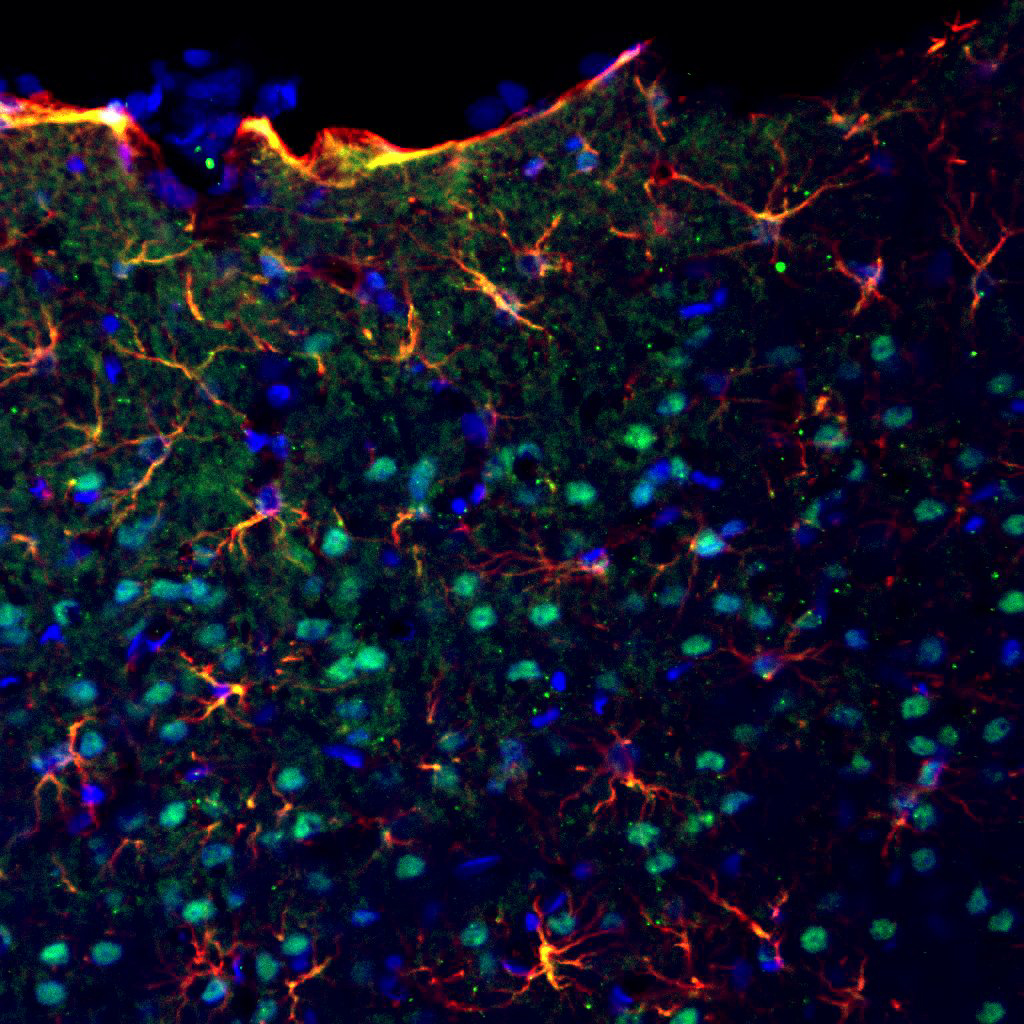
Similarly, Dr. Shai Shoham, a senior scientist at Alomone Labs, investigated MANF expression in a rat model created via intracerebroventricular (icv) streptozotocin (STZ) injection. Using IHC, Dr. Shoham demonstrated that MANF is expressed in astrocytes in a model of AD (Figure 1).
MANF Expression in Astrocytes in a Rat Model of Alzheimer’s Disease

MANF is neuroprotective in AD
Taken together, this research suggests that not only is MANF expressed in neurons and astrocytes – in AD-relevant regions of the brain – it also protects neurons against the effects of ER stress and Aβ-induced neurotoxicity. These findings offer novel insights into MANF’s role in AD pathology and its potential as both a therapeutic candidate and diagnostic marker.
Helping you explore astrocyte reactions to brain pathology
We’ve been producing and validating a range of antibodies in-house for decades. If you enjoyed the research discussed here, you might be interested in these primary antibodies:
- Anti-MANF/ARMET Antibody (#ANT-028)
- Anti-GFAP Antibody (#AFP-001) [for GFAP expression in rats]
- Anti-Connexin-43 Antibody (#ACC-201)
- Anti-Connexin-37 Antibody (#ACC-204)
- Anti-LPAR4 (P2Y9) (extracellular) Antibody (#ALR-034)
- Anti-α1-Syntrophin (SNTA1) Antibody (#APZ-021)
- Anti-SGLT1 (extracellular) Antibody (#AGT-031)
We also have blocking peptides to be used as controls for each of these antibodies. These are the original antigens we used for immunization during antibody creation:
- MANF/ARMET Blocking Peptide (#BLP-NT028)
- GFAP Blocking Peptide (#BLP-FP001)
- Connexin-43 Blocking Peptide (#BLP-CC201)
- Connexin-37 Blocking Peptide (#BLP-CC204)
- LPAR4/P2Y9 (extracellular) Blocking Peptide (#BLP-LR034)
- α1-Syntrophin/SNTA1 Blocking Peptide (#BLP-PZ021)
- SGLT1 (extracellular) Blocking Peptide (#BLP-GT031)