Overview
- Peptide CSPAPGSWLNLSHVDGN, corresponding to amino acid residues 22-38 of the rat µ-Opioid receptor (Accession P33535). Extracellular, N-terminus.

 Western blot analysis of rat hippocampus lysate:1. Anti-µ-Opioid Receptor (OPRM1) (extracellular) Antibody (#AOR-011), (1:200).
2. Anti-µ-Opioid Receptor (OPRM1) (extracellular) Antibody, preincubated with µ-Opioid Receptor/OPRM1 (extracellular) Blocking Peptide (#BLP-OR011).
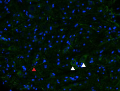
 Expression of µ-opioid receptor (MOR-1) in rat spinal cordImmunohistochemical staining of rat spinal cord frozen section using Anti-µ-Opioid Receptor (OPRM1) (extracellular) Antibody (#AOR-011), (1:100), followed by goat anti-rabbit AlexaFluor-488 secondary antibody (green). Staining is present in both neuronal cell bodies (white arrows) and their prolongations (red arrows). Hoechst 33342 is used as the counterstain (blue).
- Rat neonatal drenomedullary chromaffin cells (AMCs) (Salman, S. et al. (2013) J. Physiol. 591, 515.).
 Expression of µ-opioid receptor (MOR-1) in rat C6 cellsCell surface detection of µ-opioid receptor (MOR-1) in intact living rat brain glioma (C6) cells. A. Extracellular staining of cells using Anti-µ-Opioid Receptor (OPRM1) (extracellular) Antibody (#AOR-011), (1:300), (red). B. Merged image of A with live view of cell.
- Pan, L. et al. (2005) Neuroscience 133, 209.
- Matthes, H.W. et al. (1998) J. Neurosci. 18, 7285.
- Shaqura, M.A. et al. (2004) J. Pharmacol. Exp. Ther. 308, 712.
- Baraldi, P.G. et al. (2006) Curr. Med. Chem. 13, 3467.
Endogenous opiates such as endorphins, endomorphins, and enkephalins, as well as opiate drugs (including morphine) exert their effects by binding to opioid receptors. Three "classic" types of opioid receptors have been identified: mu (µ)-opioid (MOP) receptor, delta (δ)-opioid (DOP) receptor, and kappa (κ)-opioid (KOP) receptor. Recently, the nociceptin/orphanin FQ (N/OFQ) peptide (NOP) receptor was also described. Despite its significant sequence homology, its pharmacological profile differs greatly from those of the classic µ, δ, and κ receptors.1
The opioid receptors belong to the G protein-coupled receptor (GPCR) superfamily whose members share a common structure of seven putative transmembrane domains, an extracellular amino terminus, a cytoplasmic carboxyl terminus, and a third intracellular loop important for binding G proteins.2
All three receptors mediate opioid-induced analgesia. Supraspinal analgesia is mainly mediated by the µ-receptors, whereas µ-, δ-, and κ-receptors participate in the control of pain at the spinal level. These receptors also mediate the mood-altering properties of opioids.3
Of the opioid receptors, the µ-opioid receptor has been the most extensively studied due to its important role in mediating the actions of morphine and other analgesic agents, as well as other addictive drugs such as heroin.1 The µ-opioid receptors are expressed in the central nervous system (CNS) and in the peripherial nervous system. The highest densities are found in the thalamus, caudate putamen, neocortex, amygdala, and other brain regions known to have well established roles in pain and analgesia.4
Application key:
Species reactivity key:
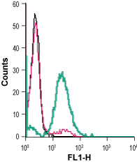
Anti-µ-Opioid Receptor (OPRM1) (extracellular) Antibody (#AOR-011) is a highly specific antibody directed against an epitope of the rat protein. The antibody can be used in western blot, immunohistochemistry, live cell imaging, and indirect live cell flow cytometry applications. It has been designed to recognize MOR-1 from rat, human, and mouse samples.

Expression of µ-Opioid Receptor in rat brain.Immunohistochemical staining of rat brain sections using Anti-µ-Opioid Receptor (OPRM1) (extracellular) Antibody (#AOR-011). MOR-1 expression is shown at the level of the anterior cingulate cortex (ACC), (A, B). Local ablation of MOR-1 staining using Derm-SAP (a cytotoxic ribosome inhibitor) abolishes MOR-1 staining (C). D, E. show higher magnification of the rectangular area of B and C, respectively.Adapted from Navratilova, E. et al. (2015) J. Neurosci. 35, 7264. with permission of the Society for Neuroscience.
Applications
Citations
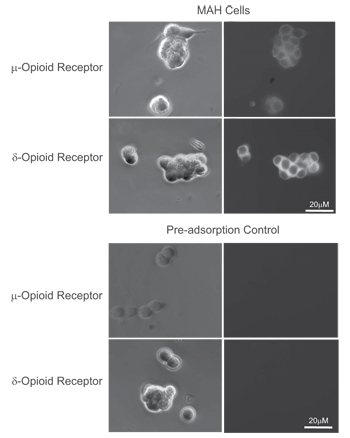
 Expression of μ- and δ-opioid receptors in immortalized rat chromaffin (MAH) cells.Immunocytochemical staining of immortalized rat chromaffin (MAH) cells. A, C. Phase contrast image of MAH cells. B, D. Staining of cells with Anti-µ-Opioid Receptor (OPRM1) (extracellular) Antibody (#AOR-011), (B) and Anti-δ-Opioid Receptor (OPRD1) (extracellular) Antibody (#AOR-014), (D). E, G. Phase contrast image of MAH cells. F, H. Anti-µ-Opioid Receptor (OPRM1) (extracellular) Antibody (F) and Anti-δ-Opioid (OPRD1) Receptor (extracellular) antibody were incubated prior to incubation with cells.
Adapted from Salman, S. et al. (2014) with permission of the American Physiological Society.
- Rat DRG lysate.
Ferrari, L.F. et al. (2019) Neuroscience 398, 64.
- Rat brain sections.
Navratilova, E. et al. (2015) J. Neurosci. 35, 7264.
- Rat immortalized chromaffin (MAH) cells.
Salman, S. et al. (2014) Am. J. Physiol. 307, C266. - Rat neonatal adrenomedullary chromaffin cells (AMCs).
Salman, S. et al. (2013) J. Physiol. 591, 515.

