Overview
- Peptide (C)KNANFTGPNQTSSNS, corresponding to amino acid residues 21-35 of human α1B-adrenoceptor (Accession P35368). Extracellular, N-terminus.
- Rat GH3 pituitary adenoma cells (10 µg/5x105 cells).
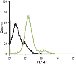 Cell surface detection of α1B-Adrenoceptor in live rat GH3 pituitary adenoma cells:___ Cells.
___ Cells + Anti-α1B-Adrenergic Receptor (extracellular)-ATTO Fluor-488 Antibody (#AAR-018-AG), (10 µg/5x105 cells).
- Rat GH3 pituitary cells.
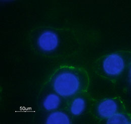 Expression of α1B-Adrenoceptor in rat GH3 cellsCell surface detection of α1B-Adrenoceptor in GH3 cells with Anti-α1B-Adrenergic Receptor (extracellular)-ATTO Fluor-488 Antibody (#AAR-018-AG) (green). Nuclear fluorescence staining of cells using the membrane-permeable DNA dye Hoechst 33342 (blue).
Adrenergic receptors (also called adrenoceptors) are the receptors for the catecholamines adrenaline and noradrenaline (called epinephrine and norepinephrine in the United States). Adrenaline and noradrenaline play important roles in the control of blood pressure, myocardial contractile rate and force, airway reactivity, and a variety of metabolic and central nervous system functions.
Adrenergic receptors are members of the G-protein coupled receptor (GPCR) superfamily of membrane proteins. They share a common structure of seven putative transmembrane domains, an extracellular amino terminus, and a cytoplasmic carboxyl terminus.
Adrenoceptors are divided into three types: α1, α2 and β adrenoceptors. Each type is further divided into at least three subtypes: α1A, α1B, α1D, α2A, α2B, α2C, β1, β2, β3.1,2 They are expressed in nearly all peripheral tissues and in the central nervous system.1,2
All α1-adrenoceptors (α1-ARs) activate phospholipases C and A2.3 In addition to mobilizing intracellular calcium, the α1-ARs have also been shown to activate calcium influx via voltage-dependent and -independent calcium channels.4
α1B-Adrenoceptor populations are greatest in the spleen, kidney, cerebellum, and fetal brain.5
α1B-Adrenoceptor causes contraction of smooth muscle cells and thereby controls vascular tone, blood pressure, and accelerates the development of atherosclerosis.5
