Overview
- Peptide (C)SEASNWTIDAENR, corresponding to amino acid residues 40-52 of rat 5-Hydroxytryptamine receptor 2A (Accession P14842). Extracellular, N-terminus.

 Western blot analysis of rat DRG (lanes 1 and 4), mouse brain (lanes 2 and 5) and human brain neuroblastoma SH-SY5Y (lanes 3 and 6) lysates:1-3. Anti-5HT2A Receptor (HTR2A) (extracellular) Antibody (#ASR-033), (1:200).
4-6. Anti-5HT2A Receptor (HTR2A) (extracellular) Antibody, preincubated with 5HT2A Receptor/HTR2A (extracellular) Blocking Peptide (#BLP-SR033).
- Mouse trigeminal ganglion sections (1:500) (Chang, W. et al. (2016) Proc. Natl. Acad. Sci. U.S.A. 113, E5491.).
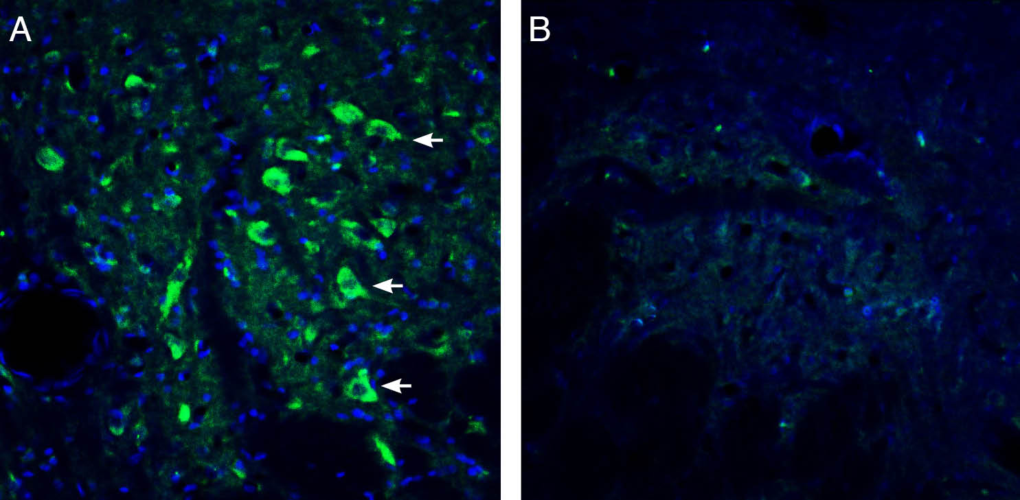
 Expression of 5HT2A Receptor in rat cingulate cortex.Immunohistochemical staining of perfusion-fixed frozen rat brain sections with Anti-5HT2A Receptor (HTR2A) (extracellular) Antibody (#ASR-033), (1:1200), followed by goat anti-rabbit-AlexaFluor-488. A. 5HT2A Receptor immunoreactivity (green) appears in outlines of cortical neurons (arrows). B. Pre-incubation of the antibody with 5HT2A Receptor (HTR2A) (extracellular) Blocking Peptide (BLP-SR033), suppressed staining. Cell nuclei are stained with DAPI (blue).
 Expression of 5HT2A Receptor in rat dorsal raphe nucleus.Immunohistochemical staining of perfusion-fixed frozen rat brain sections with Anti-5HT2A Receptor (HTR2A) (extracellular) Antibody (#ASR-033), (1:1200), followed by goat anti-rabbit-AlexaFluor-488. A. 5HT2A Receptor immunoreactivity (green) appears in outlines of neurons (arrows). B. Pre-incubation of the antibody with 5HT2A Receptor (HTR2A) (extracellular) Blocking Peptide (BLP-SR033), suppressed staining. Cell nuclei are stained with DAPI (blue).
- Hutcheson, J.D. et al. (2011) Pharmacol. Ther. 132, 146.
- Fitzgerald, L.W. et al. (2000) Mol. Pharmacol. 57, 75.
- Lorke, D.E. et al. (2006) BMC Neurosci. 7, 36.
- Raote, I. et al. (2007) Chattopadhyay A, editor. Serotonin receptors in neurobiology. Boca Raton, FL: CRC Press. 105.
5-HT is involved in a diverse array of physiological and biological processes. In the brain, 5-HT affects sleep, mood, appetite, anxiety, aggression, perception, pain, and cognition1. Signaling of 5-HT is mediated by receptors that are located on the cell membrane of neurons and most other cells in the body. The 5-HT2 family consists of three G-protein coupled receptors (GPCRs): 5-HT2A, 5-HT2B, and 5-HT2C2. They are transmembrane proteins consisting of seven membrane-spanning α-helical segments with an extracellular N-terminus and an intracellular C-terminus. The binding of 5-HT to one of its receptors is thought to elicit a conformational change that activates associated heterotrimeric G proteins and recruits other downstream signaling/scaffolding molecules, such as GPCR kinases and β-arrestins.
5-HT2A receptors have been found in many regions of the human brain including the cortex, brainstem, olfactory bulb, limbic system and basal ganglia3.
The 5-HT2A receptor has been implicated in many mental disorders including schizophrenia, depression, obsessive compulsive disorder (OCD), attention deficit–hyperactivity disorder (ADHD), and eating disorders such as anorexia nervosa and autism spectrum disorders4.
Application key:
Species reactivity key:
Alomone Labs is pleased to offer a highly specific antibody directed against an epitope of the rat 5-HT-2A receptor. Anti-5HT2A Receptor (HTR2A) (extracellular) Antibody (#ASR-033) can be used in western blot, immunohistochemistry, and live cell flow cytometry applications. The antibody recognizes an extracellular epitope and is ideal for detecting the receptor in living cells. It has been designed to recognize rat 5-hydroxytryptamine receptor 2A from rat, mouse, and human samples.

Expression of metabotropic and ionotropic 5-HT Receptors in mouse trigeminal ganglion.Immunohistochemical staining of mouse trigeminal ganglion (TG) sections using Alomone Labs Anti-5HT2A Receptor (HTR2A) (extracellular) Antibody (#ASR-033), Anti-5HT2B Receptor (HTR2B) (extracellular) Antibody (#ASR-035) and Anti-5HT3A Receptor (HTR3A) Antibody (#ASR-031) (red) shows that all three receptors are expressed in retrogradely labeled whisker afferent neurons.Adapted from Chang, W. et al. (2016) with permission of the National Academy of Sciences, USA.
Applications
Citations
- Mouse trigeminal ganglion sections (1:500).
Chang, W. et al. (2016) Proc. Natl. Acad. Sci. U.S.A. 113, E5491.

