Overview
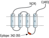
- Peptide (C)RPVPDWLRHLVLDR, corresponding to amino acid residues 342-355 of rat 5-Hydroxytryptamine receptor 3A (Accession P35563). 2nd intracellular loop.

 Western blot analysis of rat (lanes 1 and 3) and mouse (lanes 2 and 4) brain lysates:1,2. Anti-5HT3A Receptor (HTR3A) Antibody (#ASR-031), (1:200).
3,4. Anti-5HT3A Receptor (HTR3A) Antibody, preincubated with 5HT3A Receptor/HTR3A Blocking Peptide (#BLP-SR031).
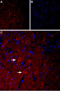
 Expression of Serotonin receptor 3A in rat brainImmunohistochemical staining of rat parietal cortex using Anti-5HT3A Receptor (HTR3A) Antibody (#ASR-031), (1:400), (red). A. 5-HT3A staining appears in neuronal outlines (arrows). B. Nuclear staining using DAPI as the counterstain. C. Merged image of A and B.
- Thompson, A.J. and Lummis, S.C. (2006) Curr. Pharm. Des. 12, 365.
- Faerber, L. et al. (2007) Eur. J. Pharmacol. 560, 1.
- Ronde, P. and Nichols, R.A. (1998) J. Neurochem. 70, 1094.
- Huang, J. et al. (2008) Neurosci. Lett. 441, 1.
5-Hydroxytryptamine receptor 3A (5-HT3A, HTR3A) belongs to the superfamily of ligand-gated ion channels. Serotonin receptors, other than the 5-HT3 subtype, belong to the superfamily of G-protein coupled receptors.
The 5-HT3 receptor is formed by five subunits arranged around a pore forming unit. Receptors could be either monomeric, such as 5-HT3A or heteromeric entities like 5-HT3A/B. Indeed, the type of channel formed displays different pharmacological and electrophysiological characteristics1,2. To date, five 5-HT3 subunits have been identified and labeled 5-HT3A-E, which show variability in the N-terminus and in the transmembrane region2. 5-HT3A and 5-HT3B are the best characterized among the different types.
In general, 5-HT3 receptors are located in the peripheral and central nervous system, in lymphocytes and intestinal enterochromaffine cells2. In presynaptic neurons, activation of these receptors leads to an increase in intracellular Ca2+ (by both influx and mobilization of intracellular stores), and modulates the release of a number of neurotransmitters and neuropeptides2. At the postsynaptic level, activation leads to membrane depolarization2,3. The 5-HT3A subunit is expressed in GABAergic and enkephalinergic neurons in the spinal dorsal horn thereby marking its possible antinociceptive effect4.
5-HT3 receptors have become important targets for which to develop treatments for irritable bowel syndrome (IBS), side effects resulting from chemotherapeutic treatment, schizophrenia and bipolar disorder1,2.
Application key:
Species reactivity key:
Anti-5HT3A Receptor (HTR3A) Antibody (#ASR-031) is a highly specific antibody directed against an epitope of rat Serotonin receptor 3A. The antibody can be used in western blot and immunohistochemistry applications. It has been designed to recognize 5-HT3A from mouse, human, and rat samples.

Expression of metabotropic and ionotropic 5-HT Receptors in mouse trigeminal ganglion.Immunohistochemical staining of mouse trigeminal ganglion (TG) sections using Alomone Labs Anti-5HT2A Receptor (HTR2A) (extracellular) Antibody (#ASR-033), Anti-5HT2B Receptor (HTR2B) (extracellular) Antibody (#ASR-035) and Anti-5HT3A Receptor (HTR3A) Antibody (#ASR-031) (red) shows that all three receptors are expressed in retrogradely labeled whisker afferent neurons.Adapted from Chang, W. et al. (2016) with permission of the National Academy of Sciences, USA.
