Overview
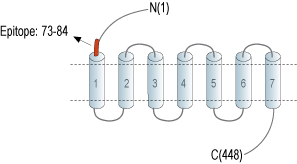
- Peptide CGEQINYGRVEK, corresponding to amino acid residues 73 - 84 of rat 5-HT7 (Accession P32305). Extracellular, N-terminus.

 Western blot analysis of rat brain lysate (lanes 1 and 3) and mouse brain lysate (lanes 2 and 4):1, 2. Anti-5HT7 Receptor/HTR7 (extracellular) Antibody (#ASR-037), (1:400).
3, 4. Anti-5HT7 Receptor/HTR7 (extracellular) Antibody, preincubated with 5HT7 Receptor/HTR7 (extracellular) Blocking Peptide (#BLP-SR037). Western blot analysis of mouse BV-2 microglia cell line lysate (lanes 1 and 4), human THP-1 monocytic leukemia cell line lysate (lanes 2 and 5) and human MEG-01 megakaryoblastic leukemia cell line lysate (lanes 3 and 6):1-3. Anti-5HT7 Receptor/HTR7 (extracellular) Antibody (#ASR-037), (1:400).
4-6. Anti-5HT7 Receptor/HTR7 (extracellular) Antibody, preincubated with 5HT7 Receptor/HTR7 (extracellular) Blocking Peptide (#BLP-SR037).
 Expression of 5HT7 Receptor in rat dorsal raphe nucleus (DRN)Immunohistochemical staining of perfusion-fixed frozen rat brain sections with Anti-5HT7 Receptor/HTR7 (extracellular) Antibody (#ASR-037), (1:300), followed by goat anti-rabbit-AlexaFluor-488. A. 5HT7R immunoreactivity (green) appears in neuronal profiles in the lateral DRN (arrows). B. Pre-incubation of the antibody with 5HT7 Receptor/HTR7 (extracellular) Blocking Peptide (#BLP-SR037), suppresses staining. Cell nuclei are stained with DAPI (blue). LDRN = lateral DRN, MDRN = medial DRN.
 Expression of 5HT7 Receptor in mouse hippocampusImmunohistochemical staining of perfusion-fixed frozen mouse brain sections with Anti-5HT7 Receptor/HTR7 (extracellular) Antibody (#ASR-037), (1:300), followed by goat anti-rabbit-AlexaFluor-488. A. Staining in the hippocampal dentate gyrus region, shows 5HT7R immunoreactivity (green) in neuronal profiles in the hilus (vertical arrows) and in glial profiles in the outer molecular layer (OML, horizontal arrows). B. Pre-incubation of the antibody with 5HT7 Receptor/HTR7 (extracellular) Blocking Peptide (#BLP-SR037), suppressed staining. Cell nuclei are stained with DAPI (blue). H = hilus, G = granule layer, OML= outer molecular layer.
- Ruat, M. et al. (1993) Proc. Natl. Acad. Sci. U.S.A. 90, 8547.
- Hedlund, P.B. and Sutcliffe, J.G. (2004) Trends Pharmacol. Sci. 25, 481.
- Vanhoenacker, P. et al. (2000) Trends Pharmacol. Sci. 21, 70.
- Thirumaran, S.L. et al. (2019) Eur. J. Med. Chem. 183, 111705.
5-HT7 receptor is a G-protein coupled receptor (GPCR) that is activated by Serotonin. Like all other members, 5-HT7 has seven transmembrane domains. In addition, it contains a hydrophobic domain located at its N-terminal end1.
The signaling cascades activated by these receptors take part in circadian rhythm, learning and memory, hippocampal signaling and smooth muscle relaxation. It was also shown to have a role in several disorders including Autism, neuropsychiatric disorders, Epilepsy and Alzheimer's disease2,4.
5-HT7 is encoded by HTR7 gene and it has three splice variants encoding proteins that differ in the length of their carboxy-terminal ends. The splice variants are differentially expressed in the spleen, kidney, heart, thalamus, hindbrain, hippocampus, cortex, caudate, striatum and cerebellum3.
Due to its involvement in central and the peripheral nervous systems, endocrine system and its involvement in various diseases and syndromes, 5-HT7 receptor represents an interesting target for the treatment and prevention of various pathologies. Selective ligands are being studied in order to regulate this receptor's activity4.
Application key:
Species reactivity key:
Anti-5HT7 Receptor/HTR7 (extracellular) Antibody (#ASR-037) is a highly specific antibody directed against an epitope of the rat protein. The antibody can be used in western blot, immunohistochemistry, and live cell flow cytometry applications. It has been designed to recognize 5HT7 from human, rat, and mouse samples.

