Overview
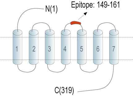
- Peptide (C)GWNRKATLASSQN, corresponding to amino acid residues 149 - 161 of mouse ADORA3 (Accession Q61618). Extracellular, 2nd loop.

 Western blot analysis of mouse P815 mastocytoma cell line lysates (lanes 1 and 3) and mouse WEHI-231 B-cell lymphoma cell line lysate (lanes 2 and 4):1-2. Anti-Adenosine A3 Receptor (extracellular) Antibody (#AAR-035), (1:200).
3-4. Anti-Adenosine A3 Receptor (extracellular) Antibody, preincubated with Adenosine A3 Receptor (extracellular) Blocking Peptide (BLP-AR035). Western blot analysis of rat spleen membranes (lanes 1 and 3) and rat brain membranes (lanes 2 and 4):1-2. Anti-Adenosine A3 Receptor (extracellular) Antibody (#AAR-035), (1:200).
3-4. Anti-Adenosine A3 Receptor (extracellular) Antibody, preincubated with Adenosine A3 Receptor (extracellular) Blocking Peptide (BLP-AR035).
- Okusa, M.D. (2002) Am. J. Physiol. Renal Physiol. 282, F10.
- Fredholm, B.B. et al. (2001) Pharmacol. Rev. 53, 527.
- Nakata, H. (1989) J. Biol. Chem. 264, 16545.
- Baraldi, P.G. et al. (2006) Curr. Med. Chem. 13, 3467.
- Headrick, J.P. et al. (2003) Am. J. Physiol. Heart Circ. Physiol. 285, H1797.
- Madi, L. et al. (2004) Clin. Cancer Res. 10, 4472.
- Yehuda.S.P. et al. (2007) Pharmacol. Rev. 16, 1601.
Adenosine is an endogenous nucleoside generated locally in tissues under conditions of hypoxia, ischemia, or inflammation. It modulates a variety of physiological functions in many tissues including the brain and heart.1,2 Adenosine exerts its action via four specific adenosine receptors (also named P1 purinergic receptors): Adenosine A1 Receptor (A1AR), Adenosine A2A Receptor (A2AAR), Adenosine A2B Receptor (A2BAR), and Adenosine A3 Receptor (A3AR). The various adenosine receptors can be distinguished on the basis of their distinct molecular structures, distinct tissue distributions, and differential selectivity for adenosine analogs.1-4 All are integral membrane proteins and are members of the G protein-coupled receptor superfamily. They share a common structure of seven putative transmembrane domains, an extracellular amino terminus, a cytoplasmic carboxyl terminus, and a third intracellular loop important in binding G proteins.1-3
Expression of A3AR has been reported in the brain, kidney, liver, and heart. It plays a role in modulation of cerebral ischemia, asthma, and cell growth. In addition, recent studies have established a cardioprotective role for A3AR.5 Expression of A3AR was reported to be elevated in cancerous tissues as well as in auto-immune inflammatory diseases.6,7 A patent has been filed for the use of A3AR and A3AR agonist as a diagnostic marker for therapeutic treatments of cancer and other diseases.
Application key:
Species reactivity key:
Anti-Adenosine A3 Receptor (extracellular) Antibody (#AAR-035) is a highly specific antibody directed against an extracellular epitope of the mouse protein. The antibody can be used in western blot and flow cytometry applications. It has been designed to recognize Adenosine A3 Receptor from rat and mouse samples. The antibody will not recognize the receptor from human samples.

