Overview
- Peptide NVGNINNDKKDETYR(C), corresponding to amino acid residues 179-193 of rat GluR2 (Accession P19491). Extracellular, N-terminus.

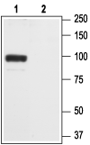
 Western blot analysis of rat brain membranes:1. Anti-GluR2 (GluA2) (extracellular) Antibody (#AGC-005), (1:200).
2. Anti-GluR2 (GluA2) (extracellular) Antibody, preincubated with GluR2/GluA2 (extracellular) Blocking Peptide (#BLP-GC005).
 Immunoprecipitation of rat brain lysates:1. Brain lysates + protein A beads + Anti-GluR2 (GluA2) (extracellular) Antibody (#AGC-005).
2. Brain lysates + protein A beads + pre-immune rabbit serum.
3. Brain lysates.
Black arrow indicates the GluR2 protein while the red arrow shows the IgG heavy chain. The immunoblot was performed with Anti-GluR2 (GluA2) (extracellular) Antibody (#AGC-005).
 Expression of AMPA Receptor 2 (GluA2) in rat cerebellumImmnunohistochemical staining of rat cerebellum using Anti-GluR2 (GluA2) (extracellular) Antibody (#AGC-005) . A. Co-localization of GluA2 (green) and calbindin D28-K (red) in Purkinje cell dendrites (vertical arrow). B. Distribution of calbindin D28-K. C. Distribution of GluA2. Note that some GluA2 positive fibers (horizontal arrow) that are probable Bergmann glial processes do not stain for GluA2. D. The molecular and Purkinje layer (blue).
Rat dissociated hippocampal neurons.
Hussain, S. and Davanger, S. (2015) PLoS ONE 10, e0140868.
- Dingledine, R. et al. (1999) Pharmacol. Rev. 51, 7.
- Song, I. and Huganir, R.L. (2002) Trends Neurosci. 25, 578.
- Malinow, R. and Malenka, R.C. (2002) Annu. Rev. Neurosci. 25, 103.
AMPA receptors are members of the glutamate receptor family of ion channels that also include the NMDA and Kainate receptors. The three subfamilies are named after the original synthetic agonists that were identified as selective ligands of each family.
The α-amino-3-hydroxy-5-methyl-4-isoazolepropionic acid (AMPA) receptor subfamily includes four members AMPA1-AMPA4 that are also known as GluR1-GluR4 respectively.
The functional AMPA channel is believed to be a tetramer, with most neuronal AMPA receptors being actually heterotetramers composed of AMPA1 plus AMPA2 or AMPA2 plus AMPA3, although homotetramers can also be found.
AMPA receptors are permeable to cations Na+, K+ and Ca2+. The Ca2+ permeability is dependent on the presence of AMPA2. The Ca2+ permeability of AMPA2 subunit is determined by the presence of the amino acid arginine (R) at a critical site in the pore loop instead of glutamine (Q) present in the same site in the other AMPA subunits. A post-transcriptional process known as RNA editing determines the presence of this R. Since most AMPA2 subunits in the adult brain have undergone RNA editing and most AMPA receptors contain the AMPA2 subunit, most native AMPA receptors will be impermeable to Ca2+.
Gating of AMPA receptors by glutamate is extremely fast and therefore the AMPA receptors mediate most excitatory (depolarizing) currents in the brain during basal neuronal activity. The depolarization caused by the activation of post-synaptic AMPA receptors is necessary for the activation of NMDA receptors that will open only in the presence of both glutamate and a depolarized membrane.
Synaptic strength, defined as the level of post-synaptic depolarization, can be long term (hence the term long term potentiation, LTP) and therefore induce changes in signaling and protein synthesis in the activated neuron. These changes are associated with memory formation and learning.
Changes in synaptic strength are thought to involve rapid movement of the AMPA receptors in and out of the synapses and a great deal of effort has been focused on understanding the mechanisms that govern AMPA receptor trafficking.
Application key:
Species reactivity key:
Anti-GluR2 (GluA2) (extracellular) Antibody (#AGC-005) is a highly specific antibody directed against an epitope of the rat ionotropic glutamate receptor 2. The antibody can be used in western blot, immunohistochemistry, live cell imaging, and immunoprecipitation applications. It has been designed to recognize GluR2 from human, mouse, and rat samples.
Applications
Citations
- Immunohistochemical staining of mouse brain sections. Tested in GLUA2-/- mice.
Egbenya, D.L. et al. (2018) Mol. Cell. Neurosci. 92, 93.
- Mouse brain lysate.
Peter, S. et al. (2020) Cell Rep. 31, 107515. - Rat brain lysate (1:500).
Szczurowska, E. et al. (2016) Exp. Neurol. 283, 97. - Rat medial prefrontal cortex (mPFC) (1:2000).
Caffino, L. et al. (2015) Addict. Biol. 20, 158. - Rat retinal lysate.
Dong, L.D. et al. (2015) J. Neurosci. 35, 5409. - Mouse brain lysate.
Khoutorsky, A. et al. (2015) eLife 4, e12002. - Rat brain lysate (1:1000).
Caffino, L. et al. (2014) Pharmacol. Rep. 66, 198. - Mouse brain lysate.
Hamada, S. et al. (2014) Eur. J. Neurosci. 40, 3136. - Rat cortical lysate (1:5000).
Banerjee, B. et al. (2013) Neurogastroenterol. Motil. 25, 973.
- Rat dissociated hippocampal neurons.
Hussain, S. and Davanger, S. (2015) PLoS ONE 10, e0140868.
- Mouse brain sections. Also tested in GLUA2-/- mice.
Egbenya, D.L. et al. (2018) Mol. Cell. Neurosci. 92, 93. - Rat brain sections.
Haglerod, C. et al. (2017) Neuroscience 344, 102. - Rat retinal sections (1:200).
Dong, L.D. et al. (2015) J. Neurosci. 35, 5409. - Rat brain sections.
Ganea, D.A. et al. (2015) Neuropsychopharmacology 40, 2727. - Fourie, C. et al. (2014) J. Neurodegener. Dis. 2014, 938530.
