Overview
- Peptide NSSTEDGIKRIQDDC, corresponding to amino acid residues 4-18 of human AT1 (Accession P30556). Extracellular, N-terminus.
- Rat kidney paraffin sections (1:50) and rat brain sections (1:60).
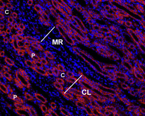
 Expression of AGTR1 in rat kidneyImmunohistochemical staining of rat kidney paraffin-embedded sections using Anti-Angiotensin II Receptor Type-1 (extracellular)-ATTO Fluor-550 Antibody (#AAR-011-AO), (1:50), (red). Staining is specific to the innermost layer of the cortex. Intense staining is present in proximal tubes (P) but not in collecting ducts (C) in the cortical labyrinths (CL). No staining is present both in thin portions of the Loop of Henle or in the collecting ducts in the medullar rays (MR). Hoechst 33342 (blue) is used to visualize the nuclei.
- Rat live intact C6 glioma cells (1:50).
Angiotensin II receptor type 1 (AT1, AGTR1) is one of the receptors that binds the octapeptide hormone Angiotensin II (Ang II).
Ang II is the peptide hormone that generates most of the known effects of the renin-angiotensin system (RAS). Ang II is generated from the angiotensinogen protein by the actions of renin, angiotensin converting enzyme (ACE) and other peptidases. Ang II has a central role in cardiovascular homeostasis by regulating vasoconstriction, renal Na+ and water readsorption. In addition, Ang II induces cell growth and proliferation and has pro-inflammatory effects.
Most of the physiological actions of Ang II are mediated by AT1 a member of the 7-transmembrane domain, G protein-coupled receptor (GPCR) superfamily.
The AT1 receptor is coupled to a Gq/11 protein that activates phospholipase C (PLC) and leads to production of inositol 1,4,5-trisphosphate (InsP3) and intracellular Ca2+ mobilization. In addition to the rapid actions mediated by InsP3 and Ca2+ signaling, AT1 receptor elicits other signaling responses including activation of the MAPK and Jak/STAT. Together, these signaling pathways mediate most of Ang II cellular responses that include, control of hormone secretion, regulation of cell growth and apoptosis, regulation of ion channels activation and cell migration.
In accordance with the pleiotropic actions of Ang II, the AT1 receptor has a wide distribution with high levels detected in the adrenal gland, kidney, brain, heart, liver, etc.
As is the case with many other peptide receptors, the AT1 receptor undergoes ligand-induced endocytosis, a process that contributes to receptor desensitization and sequestration and contributes to regulate receptor signaling.
Application key:
Species reactivity key:

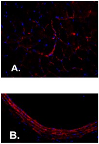
Expression of AT1 receptor in rat diaphragm and carotid body.Immunohistochemical staining of rat diaphragm muscle and carotid artery sections using Anti-Angiotensin II Receptor Type-1 (extracellular)-ATTO Fluor-550 Antibody (#AAR-011-AO). A. AT1 staining (red) in diaphragm is detected in muscle fibers. AT1 staining in carotid artery is observed in smooth muscle. DAPI is used to stain nuclei (blue).Adapted from Kwon, O.S. et al. (2015) J. Appl. Physiol. 119, 1033. with permission of the American Physiological Society.
