Overview
- Peptide NSSTEDGIKRIQDDC, corresponding to amino acid residues 4-18 of human AT1 receptor (Accession P30556). Extracellular, N-terminus.

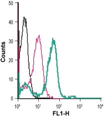
 Cell surface detection of AT1 receptor in live intact human THP-1 monocytic leukemia cells:___ Cells.
___ Cells + Rabbit IgG isotype control-FITC.
___ Cells + Angiotensin II Receptor Type-1 (extracellular)-FITC Antibody (#AAR-011-F), (5 µg). Cell surface detection of AT1 receptor in live intact human Jurkat T-cell leukemia cells:___ Cells.
___ Cells + Rabbit IgG isotype control-FITC.
___ Cells + Angiotensin II Receptor Type-1 (extracellular)-FITC Antibody (#AAR-011-F), (5 µg).
- de Gasparo, M. et al. (2000) Pharmacol. Rev. 52, 415.
- Balakumar, P. and Jagadeesh, G. (2014) J. Mol. Endocrinol. 53, R71.
- Graus-Nunes, F. et al. (2017) Mol. Cell. Endocrinol. 439, 54.
The biological functions of angiotensin II are mediated by AT1 and AT2 receptors. Both belong to the G-protein coupled receptor superfamily and are comprised of seven hydrophobic transmembrane segments forming α helices in the lipid bilayer of the cell membrane, an extracellular N-terminus and an intracellular N-terminus1.
Two highly homologous isoforms of the receptor, AT1A and AT1B have been identified in rodents. The AT1A receptor accounts for 90% of the total binding, and is predominant in the kidney, vascular smooth muscle cells, heart, liver, and in some areas of the brain, while the AT1B receptor is found predominantly in the pituitary and adrenal glands, placenta, lung, and brain. These isoforms are pharmacologically indistinguishable and are both selectively antagonized by losartan.
Through AT1 receptor, angiotensin II stimulates multiple signaling pathways, cross-talks with several tyrosine kinases, and trans-activates growth factor receptors. Phosphorylation of the receptor by GPCR kinases terminates receptor activation and promotes β-arrestin recruitment. β-arrestin-scaffolded signaling mediates ‘secondary signaling’ that involves multiple kinases that link to cell protective downstream signaling molecules2.
In a model of diet-induced obese mice, AT1 receptor inhibition demonstrates beneficial pleiotropic effects. Mice receiving the treatment exhibit enhanced pancreatic duodenal homeobox 1 (PDX1) and GLP-1 pancreatic islet expression. PDX1 islets are essential for the expression of glucokinase and GLUT2. Greater GLP-1 levels, islet vascularization and reduced apoptosis and macrophage infiltration are also observed3.
Application key:
Species reactivity key:
Anti-Angiotensin II Receptor Type-1 (extracellular) Antibody (#AAR-011) is a highly specific antibody directed against an extracellular epitope of the human protein. The antibody can be used in western blot, immunohistochemistry, immunocytochemistry, and indirect flow cytometry applications. It has been designed to recognize AT1 receptor from rat, mouse, and human samples.
Anti-Angiotensin II Receptor Type-1 (extracellular)-FITC Antibody (#AAR-011-F) is directly conjugated to fluorescein isothiocyanate (FITC). This labeled antibody can be used in immunofluorescent applications such as direct flow cytometry using live cells.
