Overview
- Peptide DNLNATGTNESAFNC, corresponding to amino acid residues 21-35 of rat AT2 receptor (Accession P35351). Extracellular, N-terminus.

 Expression of Angiotensin II receptor type-2 in rat brainImmunohistochemical staining of rat frozen free floating brain sections using Anti-Angiotensin II Receptor Type-2 (extracellular)-ATTO Fluor-488 Antibody (#AAR-012-AG), (1:20). AT2 receptor (green) was detected in neurons in the vicinity of the hypothalamic paraventricular nucleus (PVN). In some neurons (thick arrow), axonal processes with varicosities were observed (thin arrow). Nuclei were visualized with DAPI counterstain (blue).
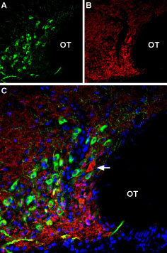
 Multiplex staining of AT2 receptor and VGLUT2 in rat supraoptic hypothalamic nucleusImmunohistochemical staining of perfusion-fixed frozen rat brain sections using Anti-Angiotensin II Receptor Type-2 (extracellular)-ATTO Fluor-488 Antibody (#AAR-012-AG), (1:60) and Anti-VGLUT2-ATTO Fluor-594 Antibody (#AGC-036-AR), (1:60). A. AT2 receptor staining (green). B. VGLUT2 staining (red). C. Merge of the two images shows a general lack of co-localization of AT2R and VGLUT2 in this part of the hypothalamus (arrow). Cell nuclei are stained with DAPI (blue).
 Expression of Angiotensin II receptor type-2 in mouse 3T3-L1 cellsCell surface detection of Angiotensin II receptor type-2 in intact live mouse 3T3-L1 cells with Anti-Angiotensin II Receptor Type-2 (extracellular)-ATTO Fluor-488 Antibody (#AAR-012-AG), (1:50), (green). Live view of the same field was superimposed to the fluorescent one.
- Mukoyama, M. et al. (1993) J. Biol. Chem. 268, 24539.
- de Gasparo, M. et al. (2000) Pharmacol. Rev. 52, 415.
- Carey, R.M. and Siragy, H.M. (2003) Endocr. Rev. 24, 261.
Angiotensin II receptor type 2 or AT2 is one of the receptors that bind the octapeptide hormone Angiotensin II (Ang II).
Ang II is the peptide hormone that generates most of the known effects of the renin-angiotensin system (RAS). Ang II is generated from the angiotensinogen protein by the actions of renin, angiotensin converting enzyme (ACE) and other peptidases. Ang II has a central role in cardiovascular homeostasis by regulating vasoconstriction, renal Na+ and water readsorption. In addition, Ang II induces cell growth and proliferation and has pro-inflammatory effects.
Most of the physiological actions of Ang II are mediated by AT1 a member of the 7-transmembrane domain, G protein-coupled receptor (GPCR) superfamily. AT2 is also a GPCR, and is largely believed to have counter-regulatory roles to the ones exerted through the AT1 receptor. Hence, binding of Ang II to the AT2 receptor will result in growth arrest and apoptosis, vasodilatation and hypotension.
Although the AT2 receptor is structurally a member of the GPCR superfamily, the signaling mechanisms elicited following AT2 receptor activation are not fully clarified. Recent evidence indicates that the AT2 receptor signals through the Giα2 and the Giα3 proteins and through the activation of phosphotyrosine phosphatases such as SHP-1.
AT2 receptor distribution is more restricted than that of the AT1 receptor. The highest expression levels of AT2 have been found in the fetus, which is followed by a marked decrease shortly after birth. Nevertheless, significant AT2 receptor expression can be detected in the heart, kidney, vascular endothelial cells and brain, among others.
Application key:
Species reactivity key:
Anti-Angiotensin II Receptor Type-2 (extracellular) Antibody (#AAR-012) is a highly specific antibody directed against an extracellular epitope of the rat protein. The antibody can be used in western blot, immunocytochemistry and immunohistochemistry applications. It has been designed to recognize AT2 receptor from rat and mouse samples. The antibody won’t recognize human AT2R.
Anti-Angiotensin II Receptor Type-2 (extracellular)-ATTO Fluor-488 Antibody (#AAR-012-AG) is directly labeled with an ATTO-488 fluorescent dye. ATTO dyes are characterized by strong absorption (high extinction coefficient), high fluorescence quantum yield, and high photo-stability. The ATTO-488 label is analogous to the well known dye fluorescein isothiocyanate (FITC) and can be used with filters typically used to detect FITC. Anti-Angiotensin II Receptor Type-2 (extracellular)-ATTO Fluor-488 Antibody has been tested in live cell imaging and immunohistochemical applications and is specially suited to experiments requiring simultaneous labeling of different markers.

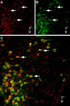
Multiplex staining of Melatonin Receptor Type 1B and AT2 Receptor in rat brainImmunohistochemical staining of perfusion-fixed frozen brain sections using Anti-Melatonin Receptor 1B (MTNR1B) Antibody (#AMR-032), (1:600) and Anti-Angiotensin II Receptor Type-2 (extracellular)-ATTO Fluor-488 Antibody (#AAR-012-AG), (1:100). A. MTNR1B staining (red) (arrows). B. The same section labeled for AT2 receptor (green). C. Merge of the two images suggests considerable co-localization in the paraventricular nucleus (arrows). For orientation, note localization with respect to 3rd ventricle (3rd V).
Applications
Citations
- Western blot analysis of mouse adrenal gland lysate with #AAR-012. Tested in AGTR2-/- mice.
Kemp, B.A. et al. (2014) Circ. Res. 115, 388.
