Overview
|
Anti-Aquaporin 3 Antibody (#AQP-003) is a highly specific antibody directed against an epitope of the rat protein. The antibody can be used in western blot, immunoprecipitation, indirect flow cytometry, and immunohistochemistry applications. It has been designed to recognize the AQP3 channel from rat, human, and mouse samples.
Application key:
Species reactivity key:

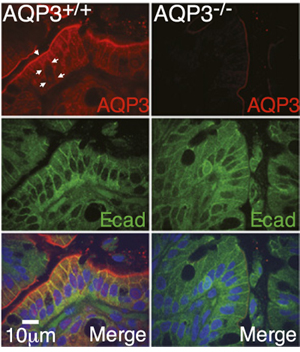
Expression of AQP3 in mouse colon.Immunohistochemical staining of mouse colon sections using Anti-Aquaporin 3 Antibody (#AQP-003). AQP3 staining (red) is detected in mouse distal colon, and co-localizes with e-cadherin (green). AQP3 is not detected in AQP3-/- cells.Adapted from Thiagarajah, J.R. et al. (2017) Proc. Natl. Acad. Sci. U.S.A. 114, 568. with permission of the National Academy of Sciences.
Applications
 Western blot analysis of rat kidney membranes:1. Anti-Aquaporin 3 Antibody (#AQP-003), (1:200).
2. Anti-Aquaporin 3 Antibody, preincubated with Aquaporin 3 Blocking Peptide (#BLP-QP003).
- Mouse epidermal keratinocytes (1 μg per ml) (Zheng, X. and Bollinger Bollag, W. et al. (2003) J. Invest. Dermatol. 121, 1487.).
 Expression of Aquaporin 3 in rat colonImmunohistochemical staining of paraffin embedded longitudinal section of rat colon using Anti-Aquaporin 3 Antibody (#AQP-003), (brown). AQP3 is detected in absorptive cells that are present in the luminal epithelium and in the superior third of the intestinal glands (arrows). Hematoxilin is used as the counterstain. Mucosa (M), submucosa (SM) and muscularis externa (ME).
- Human airway epithelial cell (Avril-Delplanque, A. et al. (2005) Stem Cells 23, 992.).
- The blocking peptide is not suitable for this application.
Citations (23)
- Immunohistochemical staining of mouse colon sections. Also tested in AQP3-/- tissue sections.
Thiagarajah, J.R. et al. (2017) Proc. Natl. Acad. Sci. U.S.A. 114, 568.
- Rat kidney lysate.
Chung, S. et al. (2019) Front. Physiol. 10, 271. - Rat digestive system lysate (1:400).
Zhao, G. et al. (2016) Biotech. Histochem. 91, 269. - Rat colon lysate.
Kon, R. et al. (2015) Toxicol. Sci. 145, 337. - Rat colon and ileum lysates (1:400).
Zhao, G. et al. (2014) Biochem. Biophys. Res. Commun. 443, 161.
- Mouse epidermal keratinocytes (1 μg per ml).
Zheng, X. and Bollinger Bollag, W. et al. (2003) J. Invest. Dermatol. 121, 1487.
- Rat kidney sections.
Chung, S. et al. (2019) Front. Physiol. 10, 271. - Mouse colon sections. Also tested in AQP3-/- tissue sections.
Thiagarajah, J.R. et al. (2017) Proc. Natl. Acad. Sci. U.S.A. 114, 568. - Sheep ovary sections (1:1000).
Sales, A.D. et al. (2016) Cell Tissue Res. 365, 415. - Rat digestive system sections (1:100).
Zhao, G. et al. (2016) Biotech. Histochem. 91, 269. - Rat colon sections.
Kon, R. et al. (2015) Toxicol. Sci. 145, 337. - Rat colon and ileum sections (1:100).
Zhao, G. et al. (2014) Biochem. Biophys. Res. Commun. 443, 161.
- Human airway epithelial cell.
Avril-Delplanque, A. et al. (2005) Stem Cells 23, 992.
- Ikarashi, N. et al. (2011) Biol. Pharm. Bull. 34, 238.
- Satake, M. et al. (2010) Biol. Pharm. Bull. 33, 1965.
- Allory, Y. et al. (2008) Kidney Int. 73, 751.
- Bae, E.H. et al. (2008) Am. J. Physiol. Renal Physiol. 294, F272.
- Ma, S.K. et al. (2007) Kidney Blood Press. Res. 30, 8.
- Leung, J.C. et al. (2005) Nephrology 10, 63.
- Edashige, K. et al. (2003) Biol. Reprod. 68, 87.
- Yeum, C.H. et al. (2003) Scand. J. Urol. Nephrol. 37, 99.
- Kim, S.W. et al. (2001) J. Am. Soc. Nephrol. 12, 875.
- Kim, S.W. et al. (2001) J. Am. Soc. Nephrol. 12, 2019.
Specifications
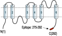
- Peptide (C)STEAENVKLAHMKHKEQI, corresponding to amino acid residues 275-292 of rat AQP3 (Accession P47862). Intracellular, C-terminus.

Scientific Background
Aquaporin 3 (AQP-3) belongs to a family of membrane proteins that allow the passage of water and certain other solutes through biological membranes. The family is composed of 13 members (AQP-0 to AQP-12).
The aquaporins can be divided into two functional groups based on their permability characteristics: the aquaporins that are only permeated by water and the aquaglyceroporins that are permeated by water and other small solutes such as glycerol. This last group includes AQP-3 as well as AQP-7, AQP-9 and AQP-10.1 Little is known about the function of the two newest members, AQP-11 and AQP-12.
The proteins present a conserved structure of six transmembrane domains with intracellular N- and C-termini. The functional channel is a tetramer but each subunit has a separate pore and therefore the functional channel unit, contains four pores.1
AQP-3 is widely expressed in several organs with prominent expression found in the skin, colon, lung and kidney. Consistent with a central function of AQP-3 in skin, mice deficient in AQP-3 have reduced skin water content and elasticity compared with wild-type mice, as well as impaired wound healing and epidermal biosynthesis.2 Furthermore, AQP-3 deficient mice were found to be resistant to skin tumor development suggesting a role for this aquaporin in tumorigenesis.3
- King, L.S. et al. (2004) Nat. Rev. Mol. Cell Biol. 5, 687.
- Hara, M. et al. (2003) Proc. Natl. Acad. Sci. U.S.A. 100, 7360.
- Hara-Chikuma, M. and Verkman, A.S. (2008) Mol. Cell. Biol. 28, 326.
