Overview
- Peptide (C)STEAENVKLAHMKHKEQI, corresponding to amino acid residues 275-292 of rat AQP3 (Accession P47862). Intracellular, C-terminus.
- Rat kidney membranes (1:200).
 Western blot analysis of rat kidney membranes:1. Anti-Aquaporin 3 Antibody (#AQP-003), (1:200).
2. Anti-Aquaporin 3 Antibody, preincubated with Aquaporin 3 Blocking Peptide (#BLP-QP003).
- Mouse epidermal keratinocytes (1 μg per ml) (Zheng, X. and Bollinger Bollag, W. et al. (2003) J. Invest. Dermatol. 121, 1487.).
- Mouse kidney sections, and rat colon.
- Human airway epithelial cell (Avril-Delplanque, A. et al. (2005) Stem Cells 23, 992.).
- The blocking peptide is not suitable for this application.
Aquaporin 3 (AQP-3) belongs to a family of membrane proteins that allow the passage of water and certain other solutes through biological membranes. The family is composed of 13 members (AQP-0 to AQP-12).
The aquaporins can be divided into two functional groups based on their permability characteristics: the aquaporins that are only permeated by water and the aquaglyceroporins that are permeated by water and other small solutes such as glycerol. This last group includes AQP-3 as well as AQP-7, AQP-9 and AQP-10.1 Little is known about the function of the two newest members, AQP-11 and AQP-12.
The proteins present a conserved structure of six transmembrane domains with intracellular N- and C-termini. The functional channel is a tetramer but each subunit has a separate pore and therefore the functional channel unit, contains four pores.1
AQP-3 is widely expressed in several organs with prominent expression found in the skin, colon, lung and kidney. Consistent with a central function of AQP-3 in skin, mice deficient in AQP-3 have reduced skin water content and elasticity compared with wild-type mice, as well as impaired wound healing and epidermal biosynthesis.2 Furthermore, AQP-3 deficient mice were found to be resistant to skin tumor development suggesting a role for this aquaporin in tumorigenesis.3
Application key:
Species reactivity key:

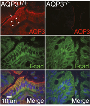
Expression of AQP3 in mouse colon.Immunohistochemical staining of mouse colon sections using Anti-Aquaporin 3 Antibody (#AQP-003). AQP3 staining (red) is detected in mouse distal colon, and co-localizes with e-cadherin (green). AQP3 is not detected in AQP3-/- cells.Adapted from Thiagarajah, J.R. et al. (2017) Proc. Natl. Acad. Sci. U.S.A. 114, 568. with permission of the National Academy of Sciences.
