Overview
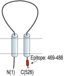
- Peptide CQKEAKRSSADKGVALSLDD, corresponding to amino acid residues 469-488 of rat ASIC1 (Accession P55926). Intracellular, C-terminus.

 Western blot analysis of rat brain membranes:1. Anti-ASIC1 Antibody (#ASC-014), (1:200).
2. Anti-ASIC1 Antibody, preincubated with ASIC1 Blocking Peptide (#BLP-SC014).
- CHO-K1 and D54-MG transfected cells (Kapoor, N. et al. (2009) J. Biol. Chem. 284, 24526.).
 Expression of ASIC1 in rat brainImmunohistochemical staining of rat globus pallidus using Anti-ASIC1 Antibody (#ASC-014). A. Parvalbumin (PV) positive neurons are shown (green). B. Neurons with typically enmeshed dendritic trees stain intensely for ASIC1 (red). C. Double labeling with mouse anti-Parvalbumin (green) reveals strong co-localization of PV with ASIC1.
- Mouse bone marrow derived macrophage (BMMs) (Kong, X. et al. (2013) Cell. Immunol. 281, 44.).
Human SH-SY5Y (Xiong, Q.J. et al. (2012) Am. J. Physiol. 303, C376.).
- Kellenberger, S. et al. (2002) Physiol. Rev. 82, 735.
- Wemmie, J.A. et al. (2002) Neuron 34, 463.
- Wemmie, J.A. et al. (2003) J. Neurosci. 23, 5496.
ASIC1 is a member of a family of Na+ channels that are activated by external protons. The family includes four additional members: ASIC2, ASIC3, ASIC4 and ASIC5. The ASICs are in fact part of a larger superfamily named degenerin/epithelial Na+ channels (DEG/ENaC) and share with it the same basic characteristics: two transmembrane spanning domains, a large extracellular domain and short intracellular N- and C-termini.
There are two recognized splice variants of the ASIC1 gene that differ on their N-termini, ASIC1a and ASIC1b that have different tissue distributions and functions.
ASIC1 responds to a decrease in extracellular pH with an inward cation current that is quickly inactivated despite the continuous presence of protons in the medium.
Recently, ASIC1 has been implicated in cognitive processes such as learning and memory.
Application key:
Species reactivity key:
Anti-ASIC1 Antibody (#ASC-014) is an antibody directed against an epitope of the rat protein. The antibody can be used in western blot, immunoprecipitation, immunocytochemistry, and immunohistochemistry applications. It has been designed to recognize ASIC1 from rat, human, and mouse samples, and recognizes both ASIC1 isoforms.

Expression of ASIC1 in rat medulla.Immunohistochemical staining of rat brain sections using Anti-ASIC1 Antibody (#ASC-014). ASIC1 staining in the medulla is detected in the ventrolateral medulla (VLM), (left panel) and in the dorsal medulla (DM), (middle panel). Antibody specificity was demonstrated by preincubating the antibody with the control antigen (right panel).Adapted from Song, N. et al. (2016) Sci. Rep. 6, 38777. with permission of SPRINGER NATURE.
Applications
Citations
- Western blot analysis of mouse lung lysate. Tested in ASIC1-/- mice.
Trac, P.T. et al. (2017) Am. J. Physiol. 312, L797.
- Mouse lung lysate. Also tested in ASIC1-/- mice.
Trac, P.T. et al. (2017) Am. J. Physiol. 312, L797. - Rat DRG lysate (1:200).
Wu, Y. et al. (2017) Cell. Mol. Neurobiol. 37, 635. - Mouse bone marrow-derived macrophage (BMM) lysate.
Kong, X. et al. (2013) Cell. Immunol. 281, 44. - Human SH-SY5Y cell lysate.
Xiong, Q.J. et al. (2012) Am. J. Physiol. 303, C376. - Mouse immature dendritic cells.
Tong, J. et al. (2011) J. Immunol. 186, 3686. - Mouse cortex lysate (1:200).
Hu, Z.L. et al. (2010) Am. J. Physiol. 299, C1355.
- CHO-K1 and D54-MG transfected cell lysate.
Kapoor, N. et al. (2009) J. Biol. Chem. 284, 24526.
- Mouse hippocampus sections (1:200).
Ferenczi, E.A. et al. (2016) Sci. Rep. 6, 23947. - Rat medulla tissue sections.
Song, N. et al. (2016) Sci. Rep. 6, 38777.
- Mouse primary DRGs (1:100).
Radu, B.M. et al. (2014) Cell Biochem. Biophys. 68, 9. - Mouse bone marrow-derived macrophages (BMMs).
Kong, X. et al. (2013) Cell. Immunol. 281, 44. - Human SH-SY5Y cell lysate.
Xiong, Q.J. et al. (2012) Am. J. Physiol. 303, C376. - Mouse dendritic cells.
Tong, J. et al. (2011) J. Immunol. 186, 3686. - Mouse cerebral cortical neurons (1:50).
Hu, Z.L. et al. (2010) Am. J. Physiol. 299, C1355.
- Zhang, Y. et al. (2012) Neurol. Sci. 33, 1125.
- Ohbuchi, T. et al. (2010) J. Physiol. 588, 2147.
- Suman, A. et al. (2010) Neurosci. Res. 68, 1.
- Yokokawa, M. et al. (2010) FEBS Lett. 584, 3107.
- Bashari, E. et al. (2009) Am. J. Physiol. 296, C372.
- Zhang, G.C. et al. (2009) Neurosci. Lett. 459, 119.
- Akiba, Y. et al. (2008) Gut 57, 1654.
- Carnally, S.M. et al. (2008) Biochem. Biophys. Res. Commun. 372, 752.
- Meltzer, R.H. et al. (2007) J. Biol. Chem. 282, 25548.
- Foulkes, T. et al. (2006) J. Neurosci. 26, 10499.
- Vila-Carriles, W.H. et al. (2006) J. Biol. Chem. 281, 19220.
- Vukicevic, M. et al. (2006) J. Biol. Chem. 281, 714.
- Donier, E. et al. (2005) J. Biol. Chem. 280, 38666.
