Overview
- Peptide DLKESPSEGSLQPSSIQC, corresponding to amino acid residues 2-18 of human ASIC2a (Accession Q16515). Intracellular, N-terminus.

 Western blot analysis of rat brain membranes:1. Anti-ASIC2a Antibody (#ASC-012), (1:200).
2. Anti-ASIC2a Antibody, preincubated with ASIC2a Blocking Peptide (#BLP-SC012).
- D54-MG transfected cell lysate (Vila Carriles, W.H. et al. (2007) J. Biol. Chem. 282, 34381.).
- Rat DRG sections (Alvarez de la Rosa, D. et al. (2002) Proc. Natl. Acad. Sci. U.S.A. 99, 2326.).
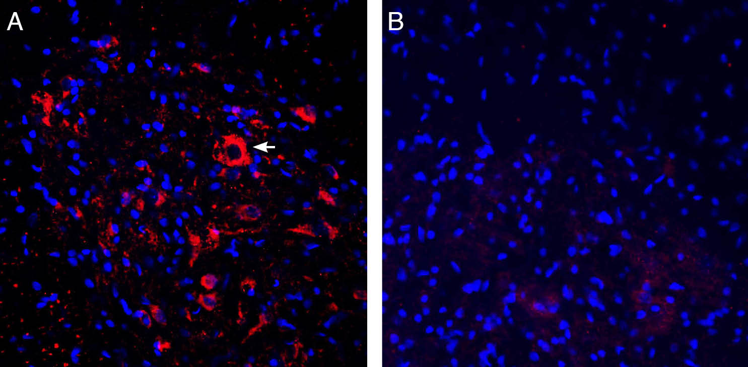
 Expression of ASIC2a in rat spinal cord.Immunohistochemical staining of perfusion-fixed frozen rat spinal cord sections with Anti-ASIC2a Antibody (#ASC-012), (1:300), followed by donkey anti-rabbit-Cy3. A. ASIC2a immunoreactivity (red) appeared in neuronal soma (horizontal arrow) in the ventral horn of the spinal cord. B. Pre-incubation of the antibody with ASIC2a Blocking Peptide (BLP-SC012), suppressed staining. Cell nuclei are stained with DAPI (blue).
- Mouse dendritic cells (Tong, J. et al. (2011) J. Immunol. 186, 3686.).
- Kellenberger, S. et al. (2002) Physiol. Rev. 82, 735.
- Krishtal, O. et al. (2003) Trends Neurosci. 26, 477.
- Price, M.P. et al. (2000) Nature 407, 1007.
ASIC2a is a member of a family of Na+ channels that are activated by external protons. The family includes four additional members: ASIC1, ASIC3, ASIC4 and ASIC5. The ASICs are in fact part of a larger superfamily named degenerin/epithelial Na+ channels (DEG/ENaC) and share with it the same basic characteristics: two transmembrane spanning domains, a large extracellular domain and short intracellular N- and C-termini.
There are two recognized splice variants of the ASIC2 gene that differ on their N-termini, ASIC2a and ASIC2b that have different tissue distributions and functions. ASIC2a is highly expressed in the central nervous system (CNS) and is less expressed in the peripheral nervous system (PNS) while ASIC2b is prominently expressed in the latter.
The functional channel is composed of 4 subunits that can be assembled as homo- or heterotetramers with the other ASIC subunits. The ASIC2b splice variant does not appear to be functional when expressed alone but it can modify the properties of ASIC2a and ASIC3 when co-expressed.
The ASIC2 protein has been proposed to be involved in mechanosensation and sensory transduction.
Application key:
Species reactivity key:
Anti-ASIC2a Antibody (#ASC-012) is a highly specific antibody directed against an epitope of the human protein. The antibody can be used in western blot, immunoprecipitation, immunohistochemistry, and immunocytochemistry applications. It has been designed to recognize ASIC2a from human, rat, and mouse samples. The antibody is specific for ASIC2a and will not recognize the ASIC2b variant.

Expression of ASIC2a in rat medulla.Immunohistochemical staining of rat brain sections using Anti-ASIC2a Antibody (#ASC-012). ASIC2a staining in the medulla is detected in the ventrolateral medulla (VLM), (left panel) and in the dorsal medulla (DM), (middle panel). Antibody specificity was demonstrated by preincubating the antibody with the blocking peptide (right panel).Adapted from Song, N. et al. (2016) Sci. Rep. 6, 38777. with permission of SPRINGER NATURE.
Applications
Citations
- Western blot analysis of human primary astrocyte lysate. Tested in siRNA-treated cells.
Vila-Carriles, W.H. et al. (2007) J. Biol. Chem. 282, 34381.
- Rat DRG lysate (1:200).
Wu, Y. et al. (2017) Cell. Mol. Neurobiol. 37, 635. - Mouse bone marrow-derived macrophage (BMM) lysate.
Kong, X. et al. (2013) Cell. Immunol. 281, 44. - Human SH-SY5Y cell lysate.
Xiong, Q.J. et al. (2012) Am. J. Physiol. 303, C376. - Mouse immature dendritic cells.
Tong, J. et al. (2011) J. Immunol. 186, 3686. - Mouse cortex lysate (1:200).
Hu, Z.L. et al. (2010) Am. J. Physiol. 299, C1355. - Mouse brain lysate.
Joch, M. et al. (2007) Mol. Biol. Cell 18, 3105. - Human primary astrocyte lysate. Also tested in siRNA-treated cells.
Vila-Carriles, W.H. et al. (2007) J. Biol. Chem. 282, 34381.
- Rat DRG sections (1:50).
Wu, Y. et al. (2017) Cell. Mol. Neurobiol. 37, 635. - Mouse hippocampus sections (1:200).
Ferenczi, E.A. et al. (2016) Sci. Rep. 6, 23947. - Rat medulla tissue sections.
Song, N. et al. (2016) Sci. Rep. 6, 38777. - Rat DRG sections.
Alvarez de la Rosa, D. et al. (2002) Proc. Natl. Acad. Sci. U.S.A. 99, 2326.
- Mouse bone marrow-derived macrophages (BMMs).
Kong, X. et al. (2013) Cell. Immunol. 281, 44. - Human SH-SY5Y cell lysate.
Xiong, Q.J. et al. (2012) Am. J. Physiol. 303, C376. - Mouse dendritic cells.
Tong, J. et al. (2011) J. Immunol. 186, 3686.
- Zhang, Y. et al. (2012) Neurol. Sci. 33, 1125.
