Overview
- Peptide (C)RQLKTVHQEYPWGE, corresponding to amino acid residues 284-297 of mouse ASIC5 (Accession Q9R0Y1). Extracellular loop.
- Rat and mouse brain lysates, rat small intestine lysate (1:200–1:1000).
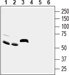
 Western blot analysis of rat brain (lanes 1 and 4), mouse brain (lanes 2 and 5) and rat small intestine (lanes 3 and 6) lysates:1-3. Anti-ASIC5 (extracellular) Antibody (#ASC-032), (1:200).
4-6. Anti-ASIC5 (extracellular) Antibody, preincubated with ASIC5 (extracellular) Blocking Peptide (#BLP-SC032).
- Mouse brain sections (1:200).
ASIC5 (acid sensing ion channel subunit family member 5) is an amiloride-sensitive ion channel belonging to the Degenerin/Epithelial Na+ channel (Deg/ENaC) family, a group of voltage-insensitive cation channels expressed in the nervous system and in several types of epithelial and immune cells.
Structurally, ASICs have two transmembrane domains which line the pore of the channel with a relatively short cytoplasmic section, intracellular N- and C-termini and a large extracellular loop. The proton ligand binds to the extracellular domain, composed of 12 β sheets and 7 α helices2,3.
ASICs are permeable to sodium, potassium, and sometimes to calcium and are activated by low pH, both intracellular and extracellular. They are responsible for conducting depolarizing inward Na+ currents. ASICs also contribute to excitotoxic neuronal damage during ischemia and local acidosis due to their sensitivity to acid1-3.
ASICs are abundantly expressed in mammalian neurons. ASIC5 is mainly expressed in the brain in the cerebellum, specifically in the ventral uvula and nodulus of the vestibulocerebellum. ASIC5 mRNA is also detected in the liver mainly in the luminal membrane of epithelial cholangiocytes that line the bile duct1.
ASICs have been linked to numerous neurological diseases including Parkinson’s disease, stroke, traumatic brain injury, multiple sclerosis, and epilepsy3.
