Overview
- Peptide (C)VLEKVPVSKGQLK, corresponding to amino acid residues 166-178 of human BDNF (precursor) (Accession P23560).

 Western blot analysis with Anti-BDNF Antibody:1,5. Recombinant human BDNF protein (#B-250).
2,6. Recombinant mouse proBDNF protein (#B-240).
3,7. Recombinant human beta-NGF protein (#N-245).
4,8. Recombinant human Neurotrophin-3 (NT-3) protein (#N-260).
Lanes 1-4: Anti-BDNF Antibody (#ANT-010), (1:200).
Lanes 5-8: Anti-BDNF Antibody, preincubated with BDNF Blocking Peptide (#BLP-NT010).
Note that the antibody recognizes both BDNF and proBDNF but fails to recognize the closely related NGF and NT-3 neurotrophins.
 Expression of BDNF in mouse cerebellumImmunohistochemical staining of mouse cerebellum with Anti-BDNF Antibody (#ANT-010). A. BDNF (red) appears in Purkinje cells (upward pointing arrows) and is distributed diffusely in the molecular layer (Mol) including in astrocytic fibers (downward pointing arrows). B. Staining of astrocytic fibers with glial fibrillary acidic protein (green) in the same section demonstrates the distribution of BDNF to neuronal as well as to astrocytic cellular components. C. Confocal merge of BDNF and GFAP.
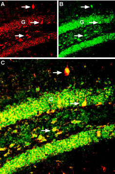
 Multiplex staining of Kir6.2 and BDNF in mouse hippocampus.Immunohistochemical staining of immersion-fixed, free floating mouse brain frozen sections using Guinea pig Anti-Kir6.2 Antibody (#APC-020-GP), (1:300) and Anti-BDNF Antibody (#ANT-010), (1:300). A. Kir6.2 staining (red) appears in the dentate gyrus granule layer (G) and in hilar interneurons (arrows). B. BDNF staining (green) in the same section appears in the dentate gyrus granule layer (G) and in hilar interneurons (arrows). C. Merge of the two images reveals colocalization of Kir6.2 and BDNF in hippocampal interneurons.
- BDNF transfected striatal cells (1:50) (del Toro, D. et al.(2006) J. Neurosci. 26, 12748.).
- Chao, M.V. (2003) Nature Rev. Neurosci. 4, 299.
- Blum, R. and Konnerth, A. (2005) Physiology 20, 70.
- Kalb, R. (2005) Trends Neurosci. 28, 5.
- Lu, B. et al. (2005) Nature Rev. Neurosci. 6, 603.
Brain-derived neurotrophic factor (BDNF) is a member of the neurotrophin family of growth factors which includes nerve growth factor (NGF), neurotrophin-3 (NT-3) and neurotrophin-4/5 (NT-4/5).
All neurotrophins are synthesized as preproneurotrophin precursors that are subsequently processed within the intracellular transport pathway to yield proneurotrophins that are further processed to generate the mature form. The mature form of BDNF is a non-covalent stable homodimer that can be secreted in both constitutive and regulated pathways.
BDNF conveys its activity by binding to two classes of receptors, a member of the Trk receptor tyrosine kinase family (TrkB) and the pan-neurotrophin receptor p75NTR. Binding of BDNF to the TrkB receptor triggers ligand-induced dimerization and autophosphorylation of tyrosine residues. This activates various signaling cascades such the MAPK, PI3K and PLCγ pathways that are involved in cell growth, survival and differentiation of neurons in the central and peripheral nervous system.
Interestingly, recent evidence suggests that BDNF may influence target cell function via ion channel modulation. Ion channel activity in the target cells can be modulated by a TrkB-mediated mechanism that has not yet been determined. BDNF is able to block both KV1.3 and AMPA-subtype glutamate ion channel currents in sensory neurons, while it can induce activation of the TRPC3 cation channel in neurons and of the NaV1.9 Na+ channel in hippocampal neurons. These newly recognized BDNF actions underlie its “rapid” neuronal functions that include changes in neuronal excitability, plasticity and synaptic transmission.
Application key:
Species reactivity key:
Anti-BDNF Antibody (#ANT-010) is a highly specific antibody directed against an epitope of the human protein. The antibody can be used in western blot, immunocytochemistry, and immunohistochemistry applications. It has been designed to recognize BDNF from rat, human, and mouse samples. The antibody is specific for BDNF; it does not crossreact with NGF, NT-3 or NT-4.

Expresssion of BDNF in mouse brain
Immunohistochemical staining of mouse brain sections using Anti-BDNF Antibody (#ANT-010). BDNF staining (purple) is expressed in astrocytes and co-localizes with GFAP (blue) but not with neurofilament-1 (NF-L), (green).Adapted from Fulmer, C.G. et al. (2014) J. Neurosci. 34, 8186. with permission of the Society for Neuroscience.
Applications
Citations
- Immunohistochemical staining of mouse nucleus of the solitary tract sections. Tested in inducible BDNF-knockout mice.
Sunc, C. et al. (2018) J. Neurosci. 38, 6873. - Immunohistochemical staining of rat brain sections. Tested in shBDNF-treated rats.
Taliaz, D. et al. (2010) Mol Psychiatry 15, 80.
- Mouse brain lysate (1:200).
Nguyen, L. et al. (2016) Neuroreport 27, 1004. - Rat brain lysate (1:1000).
Hovens, I.B. et al. (2015) Am. J. Physiol. 309, R148. - Mouse brain lysate (1:200).
Duarte-Neves, J. et al. (2015) Hum. Mol. Genet. 24, 5451. - Human SH-SY5Y and rat primary cortical neuron lysates (1:1000).
Shin, M.K. et al. (2014) Neuropharmacology 77, 414. - Mouse brain lysate (1:2000).
Shin, M.K. et al. (2014) Neurobiol. Aging. 35, 990. - Rat lamina terminalis lysate (1:200).
Clayton, S.C. et al. (2014) Am. J. Physiol. 306, R908. - Rat brain lysates (1:500).
Tian, X. et al. (2013) PLoS ONE 8, e69252.
- Mouse nucleus of the solitary tract sections. Also tested in inducible BDNF-knockout mice.
Sunc, C. et al. (2018) J. Neurosci. 38, 6873. - Mouse retina sections (1:200).
Benhar, I. et al. (2016) EMBO J. 35, 1219. - Mouse brain sections (1:200).
Wattananit, S. et al. (2016) J. Neurosci. 36, 4182. - Rat brain sections (8 µg/ml).
Louveau, A. et al. (2015) Glia 63, 2298. - Mouse brain sections.
Fulmer, C.G. et al. (2014) J. Neurosci. 34, 8186. - Rat brain sections. Also tested in shBDNF-treated rats.
Taliaz, D. et al. (2010) Mol Psychiatry 15, 80.
- BDNF transfected striatal cells (1:50).
Del Toro, D. et al. (2006) J. Neurosci. 26, 12748.
- Demel, C. et al. (2011) J. Neuroinflammation. 8, 7.
- Poblete-Naredo, I. et al. (2011) Neurochem. Int. 59, 1133.
- España, J. et al. (2010) J. Neurosci. 30, 9402.
- Kong, Li. et al. (2010) Neurobiol. Dis. 38, 446.
- Taliaz, D. et al. (2010) Mol Psychiatry 15, 80.
- Lewitus, G.M. et al. (2008) Brain Behav. Immun. 22, 1108.
- Fiszman, M.L. et al. (2005) J. Neurosci. 25, 2024.
