Overview
- Peptide (C)NPNKDYPGHEMD, corresponding to amino acid residues 259-270 of mouse Bestrophin-1 (Accession O88870). 3rd extracellular loop.

 Western blot analysis of rat lung (lanes 1 and 3) and rat eye (lanes 2 and 4) lysate:1,2. Anti-Bestrophin-1 (extracellular) Antibody (#ABC-001), (1:200).
3,4. Anti-Bestrophin-1 (extracellular) Antibody, preincubated with Bestrophin-1 (extracellular) Blocking Peptide (#BLP-BC001).
 Expression of Bestrophin-1 in rat lungImmunohistochemical staining of paraffin embedded rat lung sections using Anti-Bestrophin-1 (extracellular) Antibody (#ABC-001), (1:100). BEST1 is expressed both in the respiratory epithelium (red arrows) and in vascular smooth muscle (black arrows). Hematoxilin is used as the counterstain.
- Men, G. et al. (2004) Am. J. Ophtalmol. 137, 963.
- Petrukhin, K. et al. (1998) Nat. Genet. 19, 241.
- Hartzell, C.H. et al. (2008) Physiol. Rev. 88, 639.
- Kramer, F. et al. (2004) Cytogen. Genome Res. 105, 107.
- Stohr, H. et al. (2002) Eur. J. Hum. Genet. 10, 281.
- Park, H. et al. (2009) J. Neurosci. 29, 13063.
- Marmostein, A.D. et al. (2009) Prog Retin Eye Res. 28, 206.
- Tsunenari, T. et al. (2003) J. Biol. Chem. 278, 41114.
- Milenkovic, V.M. et al. (2007) J. Biol. Chem. 282, 1313.
- Xiao, Q. et al. (2008) J. Gen. Physiol. 132, 681.
- Chien, L.T. et al. (2006) J. Gen. Physiol. 128, 247.
- Tsunenari, T. et al. (2006) J. Gen. Physiol. 127, 749.
- Woo, D.H. et al. (2012) Cell 151, 25.
Mammalian Cl- channels can be broadly classified into four different families: voltage-dependent Cl- channels (CLCs), the cystic fibrosis transmembrane conductance regulator (CFTR), ligand-gated Cl- channels (γ-aminobutyric acid (GABA)) and glycine channels) and Ca2+-activated Cl- channels (Bestrophin and Anoctamin channels).
Bestrophins were first found by genetic linkage of human-Bestrophin-1 (hBest1) to a juvenile form of macular degeneration called Best vitelliform macular dystrophy (BVMD)1,2. BVMD is mainly electrophysiologically characterized by a decrease in the light peak and physiologically by the thinning of the retina layer which eventually leads to the loss of central vision3. To date Bestrophin 1-4 have been identified, although Bestrophin-3 and Bestrophin-4 have been observed only at the RNA level3. In addition, splice variants of some of these Ca2+-activated Cl- channels (CaCCs) have also been detected2,4,5. CaCCs are known to be involved in the regulation of olfaction, taste, phototransduction, and excitability in the nervous system. Recently, Bestrophin-1 was shown to be functionally expressed in astrocytes in both primary cell culture and in situ6. Bestrophin-1 is also detected in retina, brain, spinal cord and testes7.
Two different topologies for Bestrophin-1 have been proposed. The first, the preferred structure, proposes that six hydrophobic domains span the membrane8, while the second suggests that there are only four membrane-spanning domains9. Bestrophin-1, along with its counterparts, is activated by intracellular Ca2+. A recent study demonstrated that Bestrophin-1 indeed binds Ca2+ and by mutating specific residues, showed which amino acid residues are essential for binding Ca2+ 10, providing additional evidence that Bestrophin-1 is activated by direct binding of Ca2+ to the channel11,12.
Bestrophin-1 has been found to release glutamate from astrocytes and is located at microdomains near synapses13.
Application key:
Species reactivity key:
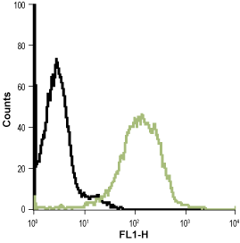
Alomone Labs is pleased to offer a highly specific antibody directed against an extracellular epitope of mouse Bestrophin-1. Anti-Bestrophin-1 (extracellular) Antibody (#ABC-001) can be used in western blot, immunohistochemistry and indirect flow cytometry applications. It has been designed to recognize bestrophin-1 channel from mouse, rat and human samples.
Applications
Citations
- Rat uterus lysate (1:2000).
Mijuškovic´, A. et al. (2015) Br. J. Pharmacol. 172, 3671. - Rat spinal cord and DRG lysates (1:200).
Pineda-Farias, J.B. et al. (2015) Mol. Pain 11, 1. - Rat DRG lysate (1:400).
Garcia, G. et al. (2014) Brain Res. 1579, 35.

