Overview
- Peptide (C)GEMMDQKDKDQVKD, corresponding to amino acid residues 22-35 of rat Betaine/GABA Transporter 1 (Accession P48056). Intracellular, N-terminus.
- Rat and mouse brain and kidney lysates (1:200-1:1000).
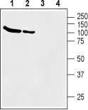
 Western blot analysis of rat (lanes 1 and 3) and mouse (lanes 2 and 4) brain membranes:1,2. Anti-BGT-1 (SLC6A12) Antibody (#AGT-004), (1:200).
3,4. Anti-BGT-1 (SLC6A12) Antibody, preincubated with BGT-1/SLC6A12 Blocking Peptide (#BLP-GT004). Western blot analysis of mouse kidney membranes:1. Anti-BGT-1 (SLC6A12) Antibody (#AGT-004), (1:200).
2. Anti-BGT-1 (SLC6A12) Antibody, preincubated with BGT-1/SLC6A12 Blocking Peptide (#BLP-GT004).
γ-aminobutyric acid (GABA) transporters (GATs) are essential regulators of the activity in the GABAergic system through their continuous uptake of the neurotransmitter from the synaptic cleft and extrasynaptic space. Four GABA transporters (GAT1, GAT2, GAT3 and BGT1) have been identified. Betaine/GABA transporter (BGT1, SLC6A12) is a member of the Na+- and Cl−-dependent neurotransmitter transporter gene family (SLC6) with a high homology to GAT1 (SLC6A1), GAT2 (SLC6A13) and GAT3 (SLC6A11). They have 12 hydrophobic transmembrane domains (TMDs), a large extracellular loop between TMD3 and 4 with multiple glycosylation sites, and intracellular N- and C-termini1,2.
BGT1 expression may vary among species. In the dog, BGT1 mRNA is primarily detected in the kidney while the levels in the brain and liver are considerably lower. The highest BGT1 mRNA levels in the mouse are in the liver, while the highest levels in man are in the kidney followed by the liver3. BGT-1 is also present in the brain, but appears to be at low levels and mostly in the leptomeninges rather than in brain tissue proper4.
Homozygous BGT1-deficient mice have normal development and show seizure susceptibility indistinguishable from that in wild-type mice in a variety of seizure threshold models5.
