Overview
- Peptide CRYNFDSSRSYDYWD, corresponding to amino acid residues 172 - 186 of mouse C3aR1(Accession O09047). Extracellular, 2nd loop.

C3aR1 (extracellular) Blocking Peptide (#BLP-AR036)
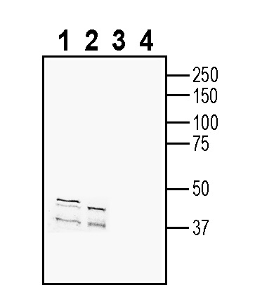
 Western blot analysis of mouse brain membranes (lanes 1 and 3) and rat brain membranes (lanes 2 and 4):1-2. Anti-C3aR1 (extracellular) Antibody (#AAR-036), (1:200).
3-4. Anti-C3aR1 (extracellular) Antibody, preincubated with C3aR1 (extracellular) Blocking Peptide (BLP-AR036). Western blot analysis of mouse M1 myeloid leukemia cell line lysate (lanes 1 and 3) and mouse J774 macrophage cell line lysate (lanes 2 and 4):1-2. Anti-C3aR1 (extracellular) Antibody (#AAR-036), (1:200).
3-4. Anti-C3aR1 (extracellular) Antibody, preincubated with C3aR1 (extracellular) Blocking Peptide (BLP-AR036).
 Expression of C3aR1 in mouse hypothalamus.Immunohistochemical staining of perfusion-fixed frozen mouse brain sections with Anti-C3aR1 (extracellular) Antibody (#AAR-036), (1:300), followed by goat anti-rabbit-AlexaFluor-488. A. Staining in the ventromedial hypothalamus shows immunoreactivity (green) in neuronal outlines (arrows). B. Pre-incubation of the antibody with C3aR1 (extracellular) Blocking Peptide (BLP-AR036), suppressed staining. Cell nuclei are stained with DAPI (blue). 3rd V = third ventricle.
 Expression of C3aR1 in rat spinal cord.Immunohistochemical staining of perfusion-fixed frozen rat spinal cord sections with Anti-C3aR1 (extracellular) Antibody (#AAR-036), (1:300), followed by goat anti-rabbit-AlexaFluor-488. A. C3aR1 immunoreactivity (green) appears in neuronal outlines (arrows). B. Pre-incubation of the antibody with C3aR1 (extracellular) Blocking Peptide (BLP-AR036), suppressed staining. Cell nuclei are stained with DAPI (blue).
- Haas, P.J. and van Srijp, J. (2007) Immun. Res. 37, 161.
- Klos, A. et al. (2009) Molec. Immun. 46, 2753.
- Gao, J. et al. (2003) J. Biol. Chem. 278, 37902.
- Gasque, P. et al. (1998) J. Immunol. 160, 3543.
- Monsinjon, T. et al. (2003) FASEB J. 17, 1003.
- Werfel, T. et al. (1997) Arch. Dermatol. Res. 289, 83.
- Hsu, M.H. et al. (1997) Immunogenetics 47, 64.
The innate immune system identifies pathogens by recognizing known elements. This response ultimately leads to the production of proinflammatory substances and the activation of phagocytic neutrophils and macrophages1. Activation of the complement system is an important initial even in the response against pathogens. This system includes some thirty different proteins that act together to form a complex1.
Three different pathways are known to activate the complement. The C3 complement plays a key role in all three pathways leading to the activation of the complement. Produced in the liver, C3 is the most abundant complement protein in the serum. It is composed of two polypeptides, an α and β chain. Cleavage of C3 leads to the production of C3b and C3a1.
C3a binds to C3a receptor, a G-protein coupled receptor which couples to Gi upon activation. Like all other members of the superfamily, C3a receptor has seven transmembrane domains with an extracellular N-terminus and intracellular C-terminal tail1. The receptor has two glycosylation sites, an unusually large extracellular loop with a sulfated tyrosine residue important for ligand recognition and binding2,3.
C3a receptor is expressed in cells of myeloid origin like neutrophils, macrophages and dendritic cells to name a few2. It is also detected in astrocytes from inflamed brain4, endothelial cells5 and activated human T cells6. mRNA of C3a receptor is detected in lung, liver, kidney, brain heart, muscle and testis2,7.
Application key:
Species reactivity key:
Anti-C3aR1 (extracellular) Antibody (#AAR-036) is a highly specific antibody directed against an extracellular epitope of the mouse protein. The antibody can be used in western blot, immunohistochemistry and flow cytometry applications. It has been designed to recognize C3aR1 from mouse and rat samples. The antibody will not recognize C3aR1 from human samples.

