Overview
- Peptide EPFPSAVTIKSWVDK(C), corresponding to amino acid residues 27-41 of rabbit CaVα2δ1 precursor (Accession P13806). Extracellular, N-terminus.

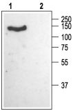
 Western blot analysis of rat brain membranes:1. Anti-CACNA2D1 (CaVα2δ1) (extracellular) Antibody (#ACC-015), (1:200).
2. Anti-CACNA2D1 (CaVα2δ1) (extracellular) Antibody, preincubated with CACNA2D1/Cavα2δ1 (extracellular) Blocking Peptide (#BLP-CC015).
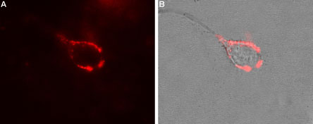
- Rat primary spinal neurons (Favereaux, A. et al. (2011) EMBO J. 30, 3830.).
- Davis, A. et al. (2007) Trends Pharm. Sci. 28, 220.
- Singer, D. et al. (1991) Science 253, 1553.
- Field, M.J. et al. (2006) Proc. Natl. Acad. Sci. U.S.A. 103, 17537.
The α2δ1 protein, encoded by CACNA2D1, is an auxiliary subunit of the CaV (voltage-dependent calcium) channel multimer complex. Four α2δ isoforms were identified in mammalian genomes. They share some sequence, as well as structure, homology.1
The protein product of the gene encoding α2δ is complex; it includes two separated proteins - the extracellular α2 peptide linked by disulfide bridges to the membrane spanning (single transmembrane helix) δ subunit.
The α2δ1 subunit is highly expressed in skeletal muscle and brain. Its expression along with a pore forming CaV α1 subunit, result in larger CaV currents, probably due to improved trafficking of the α1 subunit to the plasma membrane.1-2 α2δ1 subunit was shown to be the target of the antiepileptic and analgesic drug Gabapentin.3
Application key:
Species reactivity key:
Alomone Labs is pleased to offer a highly specific antibody directed against an epitope of rabbit CaVα2δ1. Anti-CACNA2D1 (CaVα2δ1) (extracellular) Antibody (#ACC-015) can be used in western blot and live cell imaging applications. It is especially suited to detect CaVα2δ1 in live cells. It has been designed to recognize CaVα2δ1 from rat, mouse and human samples.
Applications
Citations
- Western blot of rat hippocampal cultured cells. Tested in cells treated with alpha2delta1 shRNA.
Bikbaev, A. et al. (2020) J. Neurosci. 40, 4824. - Western blot and immunocytochemistry of mouse cells. Tested in CACNA2D1 knockout mice.
Held, R.G. et al. (2020) Neuron. 107, 667.
- Rat DRG and spinal cord lysates.
Chen, Y. et al. (2019) J. Neurochem. 148, 252. - Mouse spinal cord and small intestine lysates.
Akamine, T. et al. (2015) J. Pharmacol. Exp. Ther. 354, 65. - Transfected HEK 293 cell lysates (1:500).
Bourdin, B. et al. (2015) J. Biol. Chem. 290, 2854. - Mouse brain lysates (1:1000).
Cordeira, J.W. et al. (2014) J. Neurosci. 34, 554.
- Rat primary spinal neurons.
Favereaux, A. et al. (2011) EMBO J. 30, 3830.
- Tedeschi, A. et al. (2016) Neuron 92, 419.

