Overview
- Peptide (C)RSHSELLKKSTFAR, corresponding to amino acid residues 210-223 of rat CACNG3 (Accession Q8VHX0). Intracellular, C-terminus.
- Rat and mouse brain membranes. Human SH-SY5Y cell lysates (1:1000-1:5000).
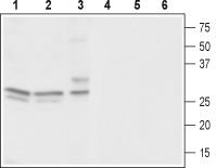
 Western blot analysis of rat brain (lanes 1 and 4), mouse brain (lanes 2 and 5) and human neuroblastoma (SH-SY5Y) cells (lanes 3 and 6):1-3. Anti-CACNG3 Antibody (#ACC-113), (1:1000).
4-6. Anti-CACNG3 Antibody, preincubated with CACNG3 Blocking Peptide (#BLP-CC113).
Voltage-gated Ca2+ (CaV) channels are ubiquitously expressed and function as Ca2+ conducting pores in the plasma membrane1. Based on their electrophysiological and pharmacological properties, Ca2+ channels have traditionally been classified into L, T, N, P/Q, and R types2.
L-type calcium channels are heteromultimers composed of four independently encoded proteins, the pore-forming α1 subunit, which triggers Ca2+ flow across the membrane, and the subunits α2δ, γ, and β3.
The γ subunit is an integral membrane protein. The γ family consists of at least 8 members, which share a number of common structural features. Each member is predicted to possess four transmembrane domains, with intracellular N- and C-termini. The first extracellular loop contains a highly conserved N-glycosylation site and a pair of conserved cysteine residues5. CaVγ subunits inhibit CaV channel activity and modulate its activation and inactivation kinetics. CaVγ subunits have little effect on CaV channel trafficking4. CaVγ3 mRNAs are only detectable in mouse brain6.
