Overview
- Peptide (C)HSLFTRSIQELDEG, corresponding to amino acid residues 19 - 32 of mouse CCR2 (Accession P51683). Extracellular, N-terminus.

CCR2 (extracellular) Blocking Peptide (#BLP-CR072)
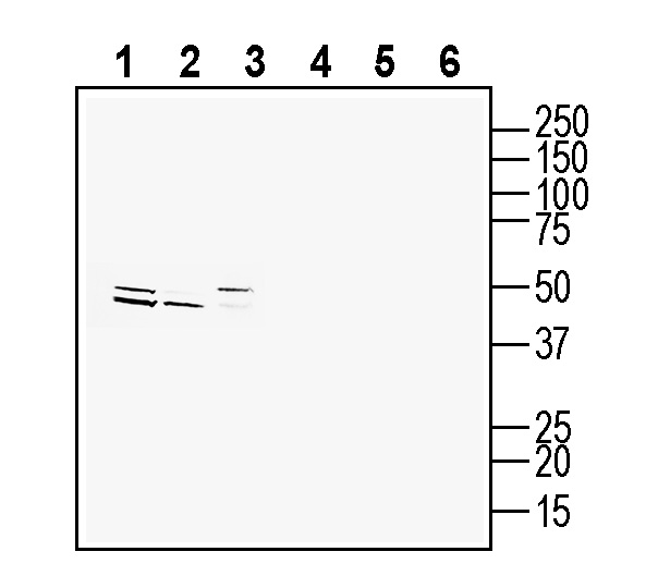
 Western blot analysis of rat brain membranes (lanes 1 and 4), mouse brain membranes (lanes 2 and 5) and mouse spleen membranes (lanes 3 and 6):1-3. Anti-CCR2 (extracellular) Antibody (#ACR-072), (1:200).
4-6. Anti-CCR2 (extracellular) Antibody, preincubated with CCR2 (extracellular) Blocking Peptide (BLP-CR072).
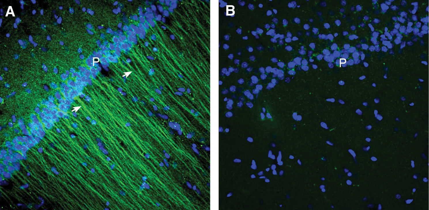
 Expression of CCR2 in mouse hippocampus.Immunohistochemical staining of perfusion-fixed frozen mouse brain sections with Anti-CCR2 (extracellular) Antibody (#ACR-072), (1:300), followed by goat anti-rabbit-AlexaFluor-488. A. CCR2 immunoreactivity (green) appears in the pyramidal layer (P) and in apical dendrites (arrows) in the hippocampal CA1 region. B. Pre-incubation of the antibody with CCR2 (extracellular) Blocking Peptide (BLP-CR072), suppressed staining. Cell nuclei are stained with DAPI (blue).
 Expression of CCR2 in rat hippocampus.Immunohistochemical staining of perfusion-fixed frozen rat brain sections with Anti-CCR2 (extracellular) Antibody (#ACR-072), (1:300), followed by goat anti-rabbit-AlexaFluor-488. A. CCR2 immunoreactivity (green) appears in the pyramidal layer (P) and in apical dendrites (arrows) in the hippocampal CA1 region. B. Pre-incubation of the antibody with CCR2 (extracellular) Blocking Peptide (BLP-CR072), suppressed staining. Cell nuclei are stained with DAPI (blue).
- Zheng, Y. et al. (2016) Nature 540, 458.
- Fantuzzi, L. et al. (2019) Cell. Mol. Life Sci. 76, 4869.
- Taylor, B.C. et al. (2019) Proc. Natl. Acad. Sci. U.S.A. 116, 8131.
- Yu, H. et al. (2022) Int. J. Mol. Sci. 23, 4642.
- Geng, H. et al. (2022) Int. J. Mol. Sci. 23, 3485.
- Curzytek, K. et al. (2021) Pharmacol. Rep. 73, 1052.
CC chemokine receptor 2 (CCR2) is one of 19 members of the chemokine receptor subfamily of class A G-protein-coupled receptors (GPCRs).
CCR2 is expressed in monocytes, immature dendritic cells, and T-cell subpopulations, and mediates acute inflammation by driving leukocyte migration to damaged or infected tissues towards chemokine ligands such as CCL2. Elevated expression of chemokines and their receptors such as CCR2 and its ligands can contribute to chronic inflammation and malignancy and hence these molecules are implicated in numerous inflammatory and neurodegenerative diseases including atherosclerosis, multiple sclerosis, asthma, neuropathic pain, and diabetic nephropathy, as well as cancer. These disease associations have motivated numerous studies in search of therapies that target the CCR2-chemokine axis1,2.
As with most G-protein coupled receptors (GPCRs), chemokine receptors transmit signals across cell membranes by means of extracellular ligand and intracellular G-protein binding. CCR2 has seven transmembrane alpha helixes, and can take distinct conformational states that are necessary for chemokine/ligand binding, G-protein binding, activation, inactivation, and signal transmission3.
CCR2 can form homo- or heterodimers with other chemokine receptors. Homodimerization may be necessary for CCR2 chemotactic activity and occurs in the absence of the ligand. CCR2/CCR5 heterocomplexes activate calcium response and support cell adhesion rather than chemotaxis, whereas CCR2/CCR4 heterodimers have an allosteric trans-inhibitory effect on CCL2 binding2.
CCR2 and its main ligand CCL2 are abundantly expressed in both the central and peripheral nervous system and have well established roles in osteoarthritis pain4, cerebral ischemia5, and depressive disorders6, among others.
Application key:
Species reactivity key:
Anti-CCR2 (extracellular) Antibody (#ACR-072) is a highly specific antibody directed against an extracellular epitope of the mouse protein. The antibody can be used in western blot, immunohistochemistry and flow cytometry applications. It has been designed to recognize CCR2 from mouse and rat samples.
The antibody will not work with human samples.

