Overview
- Peptide (C)KKAISTLTNPPAPK, corresponding to amino acid residues 674-687 of the rat longer form CLC-K2L (Accession P51802). Intracellular, C-terminus.
 Western blot analysis of rat kidney membranes:1. Anti-CLC-K Antibody (#ACL-004), (1:200).
2. Anti-CLC-K Antibody, preincubated with CLC-K Blocking Peptide (#BLP-CL004).
- Rat kidney sections.
- Jentsch, T.J. et al. (2002) Physiol. Rev. 82, 503.
- Babini, E. and Pusch, M. (2004) Physiology 19, 293.
- Estevez, R. et al. (2001) Nature 414, 558.
- Simon, D.B. et al. (1997) Nat Genet 17, 171.
CLC-Ka and CLC-Kb are members of the voltage-dependent Cl- channel (CLC) family that includes nine known members in mammals. The human CLC-Ka and CLC-Kb (known as CLC-K1 and CLC-K2 in the rat) channels are closely related genes that share 94% sequence homology and identical genomic organization.
CLC channels can be classified as plasma membrane channels and intracellular organelle channels. The first group includes the CLC-1, CLC-2, CLC-Ka and CLC-Kb channels. The second group comprises the CLC-3, CLC-4, CLC-5, CLC-6 and CLC-7.
CLC channels that function in the plasma membrane are involved in the stabilization of membrane potential and in transepithelial transport. The presumed function of the intracellular CLC channels is support of the acidification of the intraorganellar compartment. In this regard, recent reports indicate that CLC-4 and CLC-5 (and by inference CLC-3) can function as Cl-/H+ antiporters.1, 2
The functional unit of the CLC channels is a dimer with each subunit forming a proper pore. Although the crystal structure of bacterial CLC channels was resolved,the topology of the CLC channels is complex and has not been fully elucidated. It is generally accepted that both the N- and C- terminus domains are intracellular while the number and configuration of the transmembrane domains vary greatly between different models. 1,2
CLC-K channels require the presence of the auxiliary b subunit barttin, a 34 kD transmembrane protein, for transport to the plama membrane and regulation of channel permeation and gating.3
CLC-K channels are expressed primarily in the kidney from the thin ascending limb to the collecting duct of the nephron, and in the stria vascularis and dark cells of the vestibular organ of the inner ear.
The channels are important for renal salt reabsorption and water balance by enabling chloride exit across the basolateral membranes. The importance of the CLC-K channel in renal function is demonstrated by the fact that loss-of-function mutations in CLC-Kb lead to Bartter syndrome type III, an autosomal recessive disorder characterized by severe salt wasting, low blood pressure, hypokalemia and hypercalciuria.4
Application key:
Species reactivity key:
Anti-CLC-K Antibody (#ACL-004) is an antibody directed against an epitope of rat CLC-K2. The antibody can be used in western blot and immunohistochemistry applications. It has been designed to recognize CLC-K channel from rat, mouse, and human samples. The antibody recognizes both CLC-K1 and CLC-K2 isoforms.

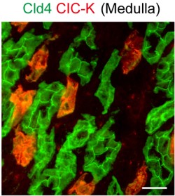
Expression of CLC-K in mouse kidney.Immunohistochemical staining of mouse kidney sections using Anti-CLC-K Antibody (#ACL-004). CLC-K staining (red) appears in the thin ascending Loop of Henle. Claudin-4 staining is shown in red.Adapted from Fujita, H. et al. (2012) PLoS ONE 7, e52272. with permission of PLoS.
Applications
Citations
- Mouse kidney sections (1:50).
Akahoshi, N. et al. (2014) Am. J. Physiol. 306, F1462. - Mouse kidney sections.
Fujita, H. et al. (2012) PLoS ONE 7, 52272.
- Tagawa, H. et al. (2005) J. Biol. Chem. 280, 23876.
- Li, W.Y. et al. (2004) Am. J. Physiol. Renal Physiol. 286, F1063.
- Kiuchi-Saishin, Y. et al. (2002) J. Am. Soc. Nephrol. 13, 875.
- Klein, J. D. et al. (2002) Am. J. Physiol. Renal Physiol. 283, F517.
- Sage, C. L. et al. (2001) Hearing Res. 160, 1.
