Overview
- Peptide (C)RNKENHKPTESSLDEK, corresponding to amino acid residues 87 - 102 of mouse CLEC7A (Accession Q6QLQ4). Extracellular, C-terminus.

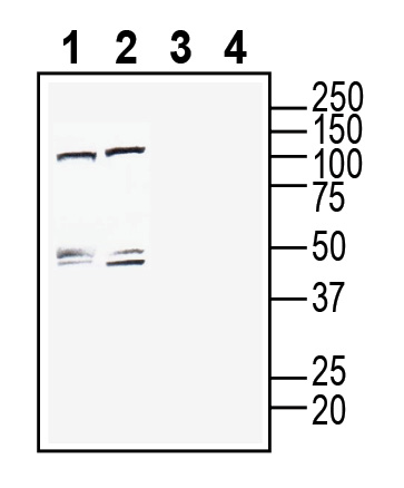
 Western blot analysis of rat brain membranes (lanes 1 and 3) and mouse brain membranes (lanes 2 and 4):1-2. Anti-CLEC7A/Dectin-1 (extracellular) Antibody (#ALR-062), (1:200).
3-4. Anti-CLEC7A/Dectin-1 (extracellular) Antibody, preincubated with CLEC7A/Dectin-1 (extracellular) Blocking Peptide (BLP-LR062). Western blot analysis of mouse BV-2 microglia cell line lysate (lanes 1 and 3) and mouse J774 macrophage cell line lysate (lanes 2 and 4):1-2. Anti-CLEC7A/Dectin-1 (extracellular) Antibody (#ALR-062), (1:200).
3-4. Anti-CLEC7A/Dectin-1 (extracellular) Antibody, preincubated with CLEC7A/Dectin-1 (extracellular) Blocking Peptide (BLP-LR062).
 Expression of CLEC7A/Dectin-1 in rat hippocampus.Immunohistochemical staining of perfusion-fixed frozen rat brain sections with Anti-CLEC7A/Dectin-1 (extracellular) Antibody (#ALR-062), (1:2400), followed by goat anti-rabbit-AlexaFluor-488. A. Staining in the hippocampal dentate gyrus region, showed CLEC7A immunoreactivity (green) in astrocytes of the hilus (H). B. Pre-incubation of the antibody with CLEC7A/Dectin-1 (extracellular) Blocking Peptide (BLP-LR062), suppressed staining. Cell nuclei are stained with DAPI (blue). G = granule layer.
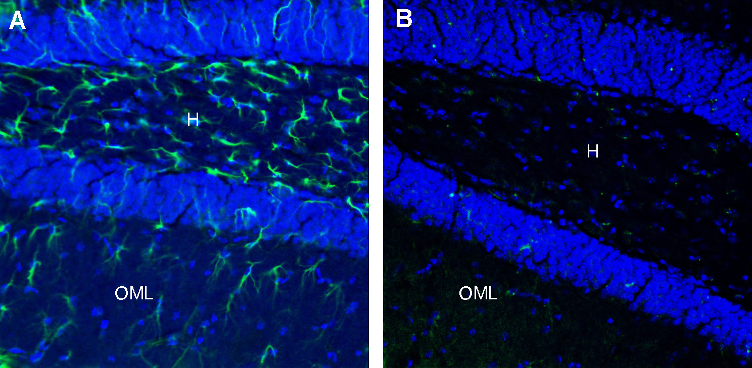
 Expression of CLEC7A/Dectin-1 in mouse hippocampus.Immunohistochemical staining of perfusion-fixed frozen mouse brain sections with Anti-CLEC7A/Dectin-1 (extracellular) Antibody (#ALR-062), (1:2400), followed by goat anti-rabbit-AlexaFluor-488. A. Staining in the hippocampal dentate gyrus region, showed CLEC7A immunoreactivity (green) in astrocytes of the hilus (H) and the outer molecular layer (OML). B. Pre-incubation of the antibody with CLEC7A/Dectin-1 (extracellular) Blocking Peptide (BLP-LR062), suppressed staining. Cell nuclei are stained with DAPI (blue).
 Multiplex staining of CLEC7A and GFAP in rat hippocampal CA1 regionImmunohistochemical staining of perfusion-fixed frozen rat brain sections with Anti-CLEC7A/Dectin-1 (extracellular) Antibody (#ALR-062), (1:600), followed by goat anti-rabbit-AlexaFluor-488 and Guinea Pig Anti-GFAP Antibody (#AFP-001-GP), (1:600), followed by goat anti-guinea pig-AlexaFluor-594. A. CLEC7A immunoreactivity (green) appears in astrocyte profiles. B. GFAP immunoreactivity (red) appears in astrocyte profiles. C. Merge of the two images demonstrates extensive co-localization. Cell nuclei are stained with DAPI (blue). P = pyramidal layer.
- Goodridge, H.S., et al., (2011) Nature, 472(7344): p. 471-5.
- Kalia, N., J. Singh, and M. Kaur, (2021) Immunobiology, 226(2): p. 152071.
- Brown, G.D., et al.,(2003) J Exp Med, 197(9): p. 1119-24.
- McDonald, J.U., et al., (2012) PLoS One, 7(9): p. e45781.
- Taylor, P.R., et al., (2002) J Immunol, 169(7): p. 3876-82.
- Kimura, Y., et al., (2014) J Biol Chem, 289(45): p. 31565-75.
- Willment, J.A., et al., (2003) J Immunol, 171(9): p. 4569-73.
- Chen, K.H., et al., (2016) Int J Biochem Cell Biol, 74: p. 135-44.
- Pamuk, O.N., et al., (2015) Exp Rheumatol, 33(4 Suppl 91): p. S15-22.
Dectin-1, encoded by CLEC7A, is a small transmembrane receptor that is a member of the C-type lectin family of pattern-recognition receptors (PRRs)1,2. As this receptor specifically recognizes and binds to β-glucans polysaccharides derived from the cell walls of fungi and yeast, it has been characterized for its role in antifungal immunity3.
Dectin-1 is predominately expressed on macrophages, dendritic cells, and neutrophils4,5. Upon ligation of β-glucan, Dectin-1 recruits the adaptor protein CARD9 to phosphorylate spleen tyrosine kinase (Syk) which in turn initiates a signaling cascade which induces phagocytosis and leads to the activation of nuclear factor-κB (NF-κB) and MAPK6. The Dectin-1 Syk mediated pathway has been demonstrated to be activated and involved in the alternative activation of macrophages as Dectin-1 was upregulated in vitro within 4 hours when stimulated with IL-4 and IL-13, cytokines which induce alternative activation of macrophages7. Moreover, the activated Syk pathway has also been implicated in renal interstitial fibrotic disease where it was demonstrated that suppressing Syk in an experimental mouse model of renal fibrosis inhibits the progression of kidney fibrosis, shown by a reduction in mRNA expression of Arginase-1 (murine M2 macrophage marker)8.
While the Dectin-1 pathway has been shown to be involved in the bleomycin-induced model of skin fibrosis, Dectin-1 signaling and receptor expression has yet to be investigated in the context of pulmonary fibrosis9.
Application key:
Species reactivity key:
Anti-CLEC7A/Dectin-1 (extracellular) Antibody (#ALR-062) is a highly specific antibody directed against an extracellular epitope of the mouse protein. The antibody can be used in western blot, immunohistochemistry and flow cytometry applications. It has been designed to recognize CLEC7A from rat and mouse samples. The antibody will not recognize CLEC7A from human samples.

