Overview
- Peptide (C)KMNGTEMNLEPGSRH, corresponding to amino acid residues 76 - 90 of mouse CNTN2 (Accession Q61330). Extracellular, N-terminus

Contactin-2/CNTN2 (extracellular) Blocking Peptide (#BLP-NR172)
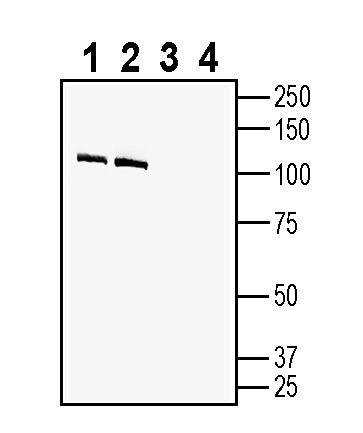
 Western blot analysis of rat brain membranes (lanes 1 and 3) and mouse brain membranes (lanes 2 and 4):1-2. Anti-Contactin-2/CNTN2 (extracellular) Antibody (#ANR-172), (1:200).
3-4. Anti-Contactin-2/CNTN2 (extracellular) Antibody, preincubated with Contactin-2/CNTN2 (extracellular) Blocking Peptide (BLP-NR172). Western blot analysis of human SH-SY5Y neuroblastoma cell line lysate:1. Anti-Contactin-2/CNTN2 (extracellular) Antibody (#ANR-172), (1:200).
2. Anti-Contactin-2/CNTN2 (extracellular) Antibody, preincubated with Contactin-2/CNTN2 (extracellular) Blocking Peptide (BLP-NR172).
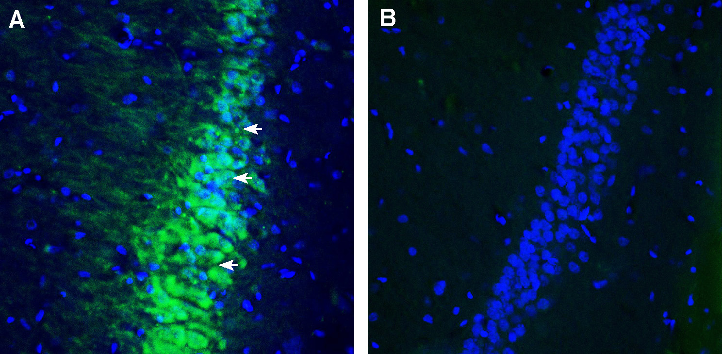
 Expression of Contactin-2 in rat hippocampus.Immunohistochemical staining of perfusion-fixed frozen rat brain sections using Anti-Contactin-2/CNTN2 (extracellular) Antibody (#ANR-172), (1:200), followed by goat anti-rabbit-AlexaFluor-488. A. Staining in the rat hippocampal CA1 region, showed Contactin-2 immunoreactivity (green) in the pyramidal layer (arrows). B. Pre-incubation of the antibody with Contactin-2/CNTN2 (extracellular) Blocking Peptide (BLP-NR172), suppressed staining. Cell nuclei are stained with DAPI (blue).
 Expression of Contactin-2 in rat cerebellum.Immunohistochemical staining of perfusion-fixed frozen rat brain sections using Anti-Contactin-2/CNTN2 (extracellular) Antibody (#ANR-172), (1:200), followed by goat anti-rabbit-AlexaFluor-488. A. Contactin-2 immunoreactivity (green) appears in purkinje cells (arrows). B. Pre-incubation of the antibody with Contactin-2/CNTN2 (extracellular) Blocking Peptide (BLP-NR172), suppressed staining. Cell nuclei are stained with DAPI (blue).
- Zinn, K. et al. (2017) Curr Opin Neurobiol. 45, 99.
- Masuda, T. et al. (2017) Cell Adh Migr. 11, 524.
- Chatterjee, M. et al. (2018) Alzheimers Res Ther. 10, 52.
- Patterson, K. R. et al. (2017) Ann Neurol. 83, 40.
- Derfuss, T. et al. (2009) Proc Natl Acad Sci USA. 106, 8302.
- Stoop, M. P. et al. (2015) Neuroimmunol Neuroinflamm. 2, 155.
Contactin-2 (also known as Axonal glycoprotein TAG-1, Axonin-1, Transient axonal glycoprotein 1, TAX-1, CNTN2) is a cell adhesion molecule that belongs to the immunoglobulin superfamily (IgSF).
IgSF proteins mediate protein-protein interactions. They are either secreted or localized to the cell surface. Many of the cell surface IgSF proteins are adhesion molecules (IgCAMs) that mediate interactions among neurons, and between neurons and glia.1
The immunoglobulin superfamily is divided into subgroups, one of which includes the contactin subfamily that consists of six members (Contactin-1 to Contactin-6). All six molecules of the contactin subgroup have six extracellular immunoglobulin-like domains and four fibronectin III-like domains, are anchored to the lipid bilayer of the cell membrane via glycosylphosphatidylinositol (GPI), and are found on the membrane surface.2
Contactin-2 is primarily expressed on the axonal and synaptic membranes. It is expressed in hippocampal pyramidal cells, cerebellar granule cells, the juxtaparanodal regions of myelinated nerve fibers, and frontal and temporal lobes. Contactin-2 is a multifunctional protein; in the embryonic nervous system, it plays important roles in axonal elongation, axonal guidance, and cellular migration. In the postnatal nervous system, it plays a role in neuronal fasciculation, axonal domain organization, formation of myelinated nerve fibers, and neuron-glia interaction.3
Contactin-2 interacts with contactin associated protein-like 2 (caspr2) at the juxtaparanodal region of nodes of Ranvier. This association is necessary for the maintenance of voltage-gated potassium channels (VGKCs) at juxtaparanodal regions of myelinated axons.
Caspr2 autoantibodies disrupt the binding of Caspr2 to contactin-2, resulting in hyperexcitability of peripheral nerves. These autoantibodies have been associated with a form of acquired peripheral nerve hyperexcitability (acquired neuromyotonia) and autoimmune encephalitis. 4
Contactin-2 is also a target protein of autoantibodies in patients with Multiple Sclerosis (MS)5. It was identified as one of the elevated cerebrospinal fluid proteins in a proteomics study in pediatric MS patients.6
Contactin-2 interacts with proteins involved in alzheimer's disease (AD) pathogenesis, such as amyloid precursor protein (APP) and beta-secretase 1 (BACE1). These interactions may influence the production of Aβ peptide and the subsequent formation of amyloid plaques. Single nucleotide polymorphisms (SNPs) in the gene encoding contactin-2 (CNTN2) are associated with AD. In addition, higher levels of contactin-2 have been reported in AD CSF pools using proteomics approaches.3
In addition, Contactin-2 is reported to be overexpressed in malignant gliomas, where it is involved in the growth and differentiation of glioma cells. It is also associated with development of oligodendrogliomas.2
Application key:
Species reactivity key:
Anti-Contactin-2/CNTN2 (extracellular) Antibody (#ANR-172) is a highly specific antibody directed against an epitope of the mouse protein. The antibody can be used in western blot and immunohistochemistry applications. It has been designed to recognize Contactin-2 from rat, mouse and human samples.
