Overview
- Peptide CNVNTHEKVKTALN, corresponding to amino acid residues 127 - 140 of human Calcitonin Receptor-Like Receptor (Accession Q16602). Extracellular, N-terminus.

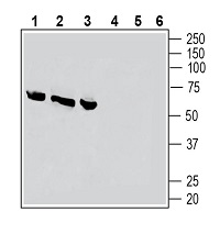
 Western blot analysis of rat dorsal root ganglion lysates (lanes 1 and 4), rat lung membranes (lanes 2 and 5) and rat heart membranes (lanes 3 and 6):1-3. Anti-CRLR/CALCRL (extracellular) Antibody (#ACR-060), (1:200).
4-6. Anti-CRLR/CALCRL (extracellular) Antibody, preincubated with CRLR/CALCRL (extracellular) Blocking Peptide (#BLP-CR060). Western blot analysis of mouse heart lysates (lanes 1 and 3) and mouse brain membranes (lanes 2 and 4):1, 2. Anti-CRLR/CALCRL (extracellular) Antibody (#ACR-060), (1:200).
3, 4. Anti-CRLR/CALCRL (extracellular) Antibody, preincubated with CRLR/CALCRL (extracellular) Blocking Peptide (#BLP-CR060). Western blot analysis of human THP-1 monocytic leukemia cell line lysate:1. Anti-CRLR/CALCRL (extracellular) Antibody (#ACR-060), (1:200).
2. Anti-CRLR/CALCRL (extracellular) Antibody, preincubated with CRLR/CALCRL (extracellular) Blocking Peptide (#BLP-CR060).
 Expression of Calcitonin Receptor-Like Receptor in rat brain stemImmunohistochemical staining of perfusion-fixed frozen rat brain sections with Anti-CRLR/CALCRL (extracellular) Antibody (#ACR-060), (1:200), followed by goat anti-rabbit-AlexaFluor-488. A. CRLR staining (green) in the Raphe Magnus region is detected in neurons (arrows). B. Pre-incubation of the antibody with CRLR/CALCRL (extracellular) Blocking Peptide (#BLP-CR060), suppresses staining. Cell nuclei are stained with DAPI (blue).
 Expression of Calcitonin Receptor-Like Receptor in mouse brain stemImmunohistochemical staining of perfusion-fixed frozen mouse brain sections with Anti-CRLR/CALCRL (extracellular) (extracellular) Antibody (#ACR-060), (1:200), followed by goat anti-rabbit-AlexaFluor-488. A. CRLR staining (green) in the Raphe Magnus region is detected in (green) in neurons (arrows). B. Pre-incubation of the antibody with CRLR/CALCRL (extracellular) Blocking Peptide (#BLP-CR060), suppresses staining. Cell nuclei are stained with DAPI (blue).
- Singh, Y. et al. (2017) CNS Neurosci. Ther. 23, 457.
- Conner, A.C. et al. (2002) Biochem. Soc. Trans. 30, 451.
- Holland, P.R. and Goadsby, P.J. (2018) Neurotherapeutics 15, 304.
Calcitonin Receptor-Like Receptor 1 (CRLR or CGRPR) is a G protein-coupled receptor that binds the peptide hormone calcitonin and is involved in maintenance of calcium homeostasis, particularly with respect to bone formation and metabolism1.
When engaged with its substrate, calcitonin gene related peptide (CGRP), CGRPR activates cAMP-dependent pkA and PI3 kinase, which eventually leads to complex inhibitory as well as facilitator actions on Neuronal Nicotinic Acetylcholine Receptor1.
CGRP receptor is a heterodimer composed of two proteins: a 7‐transmembrane domain type 2 G-protein coupled receptor called calcitonin-receptor-like receptor (CRLR), and receptor-activity-modifying protein 1 (RAMP1), a single-membrane-pass protein. RAMP1 serves as a transporter for CRLR to the cell surface and its absence leads to failure of binding with CRLR2.
CGRP and CGRPR are ubiquitously expressed and their interaction is related to various diseases and health states such as migraines3 and Alzheimer's disease1.
Application key:
Species reactivity key:
Anti-CRLR/CALCRL (extracellular) Antibody (#ACR-060) is a highly specific antibody directed against an epitope of the human protein. The antibody can be used in western blot, immunohistochemistry and indirect live cell flow cytometry. It has been designed to recognize CRLR from human, rat, and mouse samples.

