Overview
- Peptide (C)ELDLENFEYDDSAE, corresponding to amino acid residues 7- 20 of mouse CX3CR1 (Accession Q9Z0D9). Extracellular, N-terminus.

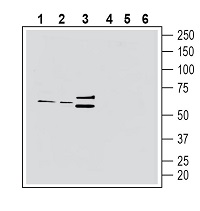
 Western blot analysis of rat brain lysate (lanes 1 and 4), mouse brain membranes (lanes 2 and 5) and rat spleen lysate (lanes 3 and 6):1-3. Anti-CX3CR1 (extracellular) Antibody (#ACR-058), (1:200).
4-6. Anti-CX3CR1 (extracellular) Antibody, preincubated with CX3CR1 (extracellular) Blocking Peptide (#BLP-CR058). Western blot analysis of mouse BV-2 microglia cell line lysate (lanes 1 and 3) and mouse J774 macrophage cell line lysate (lanes 2 and 4):1, 2. Anti-CX3CR1 (extracellular) Antibody (#ACR-058), (1:200).
3, 4. Anti-CX3CR1 (extracellular) Antibody, preincubated with CX3CR1 (extracellular) Blocking Peptide (#BLP-CR058).
 Expression of CX3CR1 in rat hippocampusImmunohistochemical staining of perfusion-fixed frozen rat brain sections with Anti-CX3CR1 (extracellular) Antibody (#ACR-058), (1:300), followed by goat anti-rabbit-AlexaFluor-488. A. CX3CR1 staining (green) in the hippocampal CA1 region is detected in microglial profiles in the stratum oriens. Some profiles include the entire microglial outline including soma (horizontal arrows) while other outlines are processes (vertical arrows). B. Pre-incubation of the antibody with CX3CR1 (extracellular) Blocking Peptide (#BLP-CR058), suppresses staining. Cell nuclei are stained with DAPI (blue). P = pyramidal layer, SR = stratum radiatum, SO = stratum oriens.
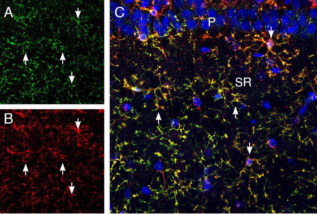
 Multiplex staining of CX3CR1 and CD11b in rat hippocampus.Immunohistochemical staining of perfusion-fixed frozen rat brain sections Anti-CX3CR1 (extracellular) Antibody (#ACR-058), (1:300), followed by goat anti-rabbit-AlexaFluor-488 and mouse Anti -CD11b, (1:500), followed by goat anti-mouse-biotin and streptavidin-Cy3. A. CXCR1 immunoreactivity (green) appears in microglial processes (up pointing arrows) and in full microglial outline (down pointing arrows). B. CD11b immunoreactivity (red) appears in microglial profiles. C. Merge of the two images demonstrates ubiquitous co-localization. Cell nuclei are stained with DAPI (blue). P = pyramidal layer, SR = stratum radiatum.
- Bronger, H. et al. (2019) Cancer Metastasis Rev. 38, 417.
- Hamon, P. et al. (2017) Blood 129, 1296.
- Luo, P. et al. (2019) Brain. Res. Bull. 146, 12.
- Lee, M. et al. (2018) Immune Netw. 18, e5.
- Asai, H. et al. (2015) Nat. Neurosci. 18, 1584.
CX3C-chemokine receptor 1 (CX3CR1) is a 368 amino acid (aa) protein that contains seven-transmembrane domains and belongs to the family of G protein coupled receptors1.
CX3CR1 has a central role in adhesion and chemotaxis through binding to its ligand, fractalkine CX3C chemokine ligand 1, (CX3CL1). The major role of CX3CR1 in immune cells is to recognize and enter inflamed tissue according to CX3CL1 gradient and initiate the innate immune system2. The receptor-ligand axis also mediates the crosstalk between glial cells and neurons by direct or indirect ways in the central nervous system (CNS)3. Expression and interaction of CX3CR1 with CX3CL1 is related to many diseases among which are asthma, cancer and gut infections (reviewed in 4).
Both CX3CL1 and CX3CR1 are expressed throughout the body. However, their expression is highly cell type-specific depending on organs and tissues. In the brain, CX3CL1/CX3CR1 signaling can modulate the production of cytokines by microglia cells. It has been reported that CX3CL1/CX3CR1 signaling is associated with Alzheimer's Disease5. In the liver, CX3CR1 is expressed in monocytes, CD8+ T cells, and natural killer (NK) cells. Besides immune cells, it is highly expressed at inflammatory sites4.
Application key:
Species reactivity key:
Anti-CX3CR1 (extracellular) Antibody (#ACR-058) is a highly specific antibody directed against an epitope of the mouse receptor. The antibody can be used in western blot, immunohistochemistry, and indirect flow cytometry applications. It has been designed to recognize CX3CR1 from mouse and rat samples. The antibody will not recognize the protein from human samples.

