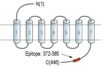
Overview
- Peptide SSHHEPRGSISKDC, corresponding to amino acid residues 372-385 of rat DRD1 (Accession P18901). Intracellular, C-terminus.

 Western blot analysis of rat brain synaptosomal fraction (lanes 1 and 3) and mouse brain synaptosomal fraction (lanes 2 and 4):1,2. Guinea pig Anti-D1 Dopamine Receptor Antibody (#ADR-001-GP), (1:400).
3,4. Guinea pig Anti-D1 Dopamine Receptor Antibody, preincubated with D1 Dopamine Receptor Blocking Peptide (#BLP-DR001). Western blot analysis of human SH-SY5Y neuroblastoma cell lysate:
1. Guinea pig Anti-D1 Dopamine Receptor Antibody (#ADR-001-GP), (1:400).
2. Guinea pig Anti-D1 Dopamine Receptor Antibody, preincubated with D1 Dopamine Receptor Blocking Peptide (#BLP-DR001).
 Expression of DRD1 in rat cortexImmunohistochemical staining of perfusion-fixed frozen rat brain sections using Guinea pig Anti-D1 Dopamine Receptor Antibody (#ADR-001-GP), (1:300), followed by goat-anti-guinea pig-Cy3 antibody. DRD1 staining (red) appears in neuronal soma (arrows). Nuclei are stained with DAPI (blue).
- Missale, C. et al. (1998) Physiol. Rev. 78, 189.
- Zeng, C. et al. (2008) Am. J. Physiol. 294, H551.
The D1 dopamine receptor (D1 receptor, DRD1) is one of five receptors that mediate the effects of the catecholamine neurotransmitter dopamine. Dopamine regulates a variety of functions including locomotor activity, emotion, positive reinforcement, food intake, and hormone secretion. The dopaminergic system has been extensively studied in the last thirty years mainly because its dysregulation has been linked to several neurological and neuropsychiatric diseases including Parkinson’s disease and Schizophrenia1.
All five dopamine receptors belong to the 7-transmembrane domain, G-protein coupled receptor (GPCR) superfamily.
Historically, the five receptors have been divided into two subfamilies based on pharmacological and structural considerations: the D1-like subfamily (that includes the D1 and D5 subtypes) and the D2-like subfamily (that includes the D2-, D3- and D4 subtypes)1.
The D1-like receptors are coupled to Gs-type G proteins and enhance adenylate cyclase activity while the D2-like receptors are coupled to Gi-type G proteins and inhibit adenylate cyclase activity1.
The D1 receptor is widely distributed throughout the brain with the highest expression in the cerebral cortex and striatum. In the periphery the D1 receptor has been detected in the adrenal cortex, kidney and heart.
Functionally, the D1 receptor has been implicated in the regulation of both locomotor and cognitive functions including the maintaining of spontaneous motor behaviors, the control of working memory and cognition as well as the regulation of craving and reward pathways.
In addition, the D1 receptor plays an important role in the pathogenesis of hypertension by regulating epithelial Na+ transport and by interacting with vasoactive hormones/humoral factors, such as aldosterone and angiotensin1,2.
Application key:
Species reactivity key:
Alomone Labs is pleased to offer a highly specific antibody directed against an epitope of rat D1 Receptor. Guinea pig Anti-D1 Dopamine Receptor Antibody (#ADR-001-GP, formerly AGP-100) raised in guinea pigs can be used in western blot and immunohistochemistry applications. It has been designed to recognize DRD1 from rat, human and mouse samples. The antigen used to immunize guinea pigs is the same as Anti-D1 Dopamine Receptor Antibody (#ADR-001) raised in rabbit. Our line of guinea pig antibodies enables more flexibility with our products such as multiplex staining studies, immunoprecipitation, etc.
Applications
Citations
- Rat spinal cord sections (1:75).
Rodgers, H.M. et al. (2019) Neuroscience 406, 376.
- Rat spinal cord sections (1:75).
Rodgers, H.M. et al. (2019) Neuroscience 406, 376.
