Overview
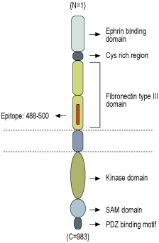
- Peptide (C)RARGTNVTISSLK, corresponding to amino acid residues 488-500 of human EphA3 (Accession P29320). Extracellular, N-terminus.

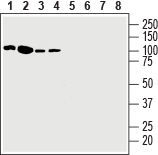
 Western blot analysis of mouse (lanes 1 and 3) and rat (lanes 2 and 4) brain membranes:1,2. Anti-EphA3 (extracellular) Antibody (#AER-013), (1:200).
3,4. Anti-EphA3 (extracellular) Antibody, preincubated with EphA3 (extracellular) Blocking Peptide (#BLP-ER013). Western blot analysis of human Jurkat T-cell leukemia cell line lysate (lanes 1 and 5), human K562 chronic myelogenous leukemia cell line lysate (lanes 2 and 6), human Malme-3M melanoma cell line lysate (lanes 3 and 7) and human HT-29 colorectal adenocarcinoma cell line lysate (lanes 4 and 8):1-4. Anti-EphA3 (extracellular) Antibody (#AER-013), (1:200).
5-8. Anti-EphA3 (extracellular) Antibody, preincubated with EphA3 (extracellular) Blocking Peptide (#BLP-ER013).
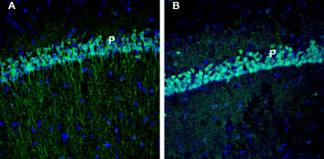
 Expression of EphA3 in rat and mouse hippocampusImmunohistochemical staining of perfusion-fixed frozen rat and mouse brain sections with Anti-EphA3 (extracellular) Antibody (#AER-013), (1:200), followed by goat anti-rabbit-AlexaFluor-488. A. EphA3 staining (green) in rat hippocampal CA1 region is detected in the pyramidal layer (P). B. In mouse hippocampal CA1 region, EphA3 immunoreactivity (green) is observed in the pyramidal layer (P). Cell nuclei are stained with DAPI (blue).
- Offenhäuser, C. et al. (2018) Cancers (Basel) 10, 12.
- Forse, G.J. et al. (2015) PLoS One 10, e0127081.
- Nasri, B. et al. (2017) BMC Clin. Pathol. 17, 8.
- Andretta, E. et al. (2017) Sci. Rep. 7, 41576.
Eph receptors are the largest family of receptor tyrosine kinases (RTKs). Based on sequence similarity and the type of ephrin ligands that they bind, they are classified into A and B sub-classes. EphA receptors bind ephrin-A ligand while EphB bind ephrin-B ligand.
Eph receptors play an important role during embryonic development and are involved in several processes including cell adhesion, cell migration, and tissue boundary formation. Eph receptors are typically expressed at low levels in normal adult tissues1,2.
EphA3 (Ephrin type-A receptor 3) structure includes a ligand-binding domain (LBD) at the N-terminus followed by a cysteine-rich region and two fibronectin III repeats. The receptor-binding domain of the ephrins is connected through a short linker segment to a GPI-anchor or a transmembrane segment3.
EphA3 was first identified as a surface antigen on pre-B lymphoblastic leukemia cells. High EphA3 levels have been detected in a number of hematological cancers and solid tumors such as lung cancers, melanomas, gastric carcinoma, leukemia, in B and T cell malignancies and in glioblastoma multiforme. In addition, high levels of EphA3 are implicated in lymph node metastasis, advanced stages of colorectal cancer, and prostate cancer1,3,4.
Application key:
Species reactivity key:
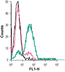
Anti-EphA3 (extracellular) Antibody (#AER-013) is a highly specific antibody directed against an epitope of the human protein. The antibody can be used in western blot, immunohistochemistry and live cell flow cytometry applications. It has been designed to recognize EphA3 from human, rat, and mouse samples.

