Overview
- Peptide (C)ERSYRIVRTAARNTD, corresponding to amino acid residues 485 - 499 of human EPHA4 (Accession P54764). Extracellular, N-terminus.
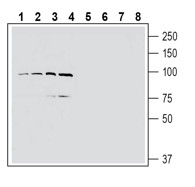
 Western blot analysis of mouse brain membranes (lanes 1 and 3) and rat brain membranes (lanes 2 and 4):1-2. Anti-EphA4 (extracellular) Antibody (#AER-044), (1:200).
3-4. Anti-EphA4 (extracellular) Antibody, preincubated with EphA4 (extracellular) Blocking Peptide (BLP-ER044). Western blot analysis of human MCF-7 breast adenocarcinoma cell line lysate (lanes 1 and 5), human U-87 MG glioblastoma cell line lysate (lanes 2 and 6), human Jurkat T-cell leukemia cell line lysate (lanes 3 and 7) and human THP-1 monocytic leukemia cell line lysate (lanes 4 and 8):1-4. Anti-EphA4 (extracellular) Antibody (#AER-044), (1:200).
5-8. Anti-EphA4 (extracellular) Antibody, preincubated with EphA4 (extracellular) Blocking Peptide (BLP-ER044).
EphA4 is a member of the Eph receptor tyrosine kinase family.
Eph receptors are subdivided into two subclasses, termed EphA and EphB, based on sequence similarity and their preference for binding a particular subclass of ephrins. The domain organization of Eph receptors contains a globular ligand binding domain (LBD), a cysteine-rich region, and two fibronectin type III domains (FN1 and FN2). It also contains a transmembrane (TM) helix, and an intracellular part consisting of a juxtamembrane (JM) region and a tyrosine kinase domain. Eph kinase activity is auto-inhibited through interaction with its own JM region, this auto inhibition is released by phosphorylation1.
Interactions of Eph receptors with their ligands, ephrins, at cell-cell interfaces promote a variety of cellular responses, including repulsion, attraction and migration2.
EphA4 is activated by all members of the ephrin-A ligand family and most members of the ephrin-B family1. Upon ligand binding it activates pathways that regulate the formation of the axon tract and cortical network as well as radial migration of cortical neurons in the cortex3.
EphA4 is expressed in the neural ectoderm at embryonic day 8 and then in various cell types throughout the cortex during subsequent stages of neurodevelopment4, its major effect on cell-cell interfaces makes this receptor highly important for the study of cellular regeneration and various types of disease.
