Overview
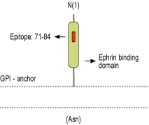
- Peptide (C)HYGAPLPPAERMER, corresponding to amino acid residues 71-84 of mouse Ephrin-A2 (Accession P52801). Extracellular.

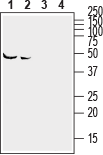
 Western blot analysis of mouse (lanes 1 and 3) and rat (lanes 2 and 4) brain lysates:1,2. Anti-Ephrin-A2 (extracellular) Antibody (#AER-032), (1:400).
3,4. Anti-Ephrin-A2 (extracellular) Antibody, preincubated with Ephrin-A2 (extracellular) Blocking Peptide (#BLP-ER032).
 Expression of Ephrin-A2 in mouse hippocampusImmunohistochemical staining of perfusion-fixed frozen mouse brain sections with Anti-Ephrin-A2 (extracellular) Antibody (#AER-032), (1:200), followed by goat-anti-rabbit-AlexaFluor-488. Ephrin-A2 staining (green) in hippocampal dentate gyrus appears in neurons (vertical arrows) and astrocytes (horizontal arrows). Cell nuclei are stained with DAPI (blue).
- Homman-Ludiye, J. et al. (2017) Sci. Rep. 7, 11813.
- Mukai, M. et al. (2017) BMC Cell Biol. 18, 28.
- Xu, Q. et al. (2000) Philos. Trans. R. Soc. Lond. B Biol. Sci. 355, 993.
Eph receptor tyrosine kinases family and their ligands, ephrins, are membrane proteins that primarily regulate cell to cell repulsion and cell adhesion and movement by modulating the organization of the actin cytoskeleton mainly through Rho family GTPases. Eph receptors and ephrin ligand signaling regulaes many aspects of cell interaction in the developing neocortex, including migration and axonal target recognition. Ephrin signaling plays an important role in guiding neurons and their projections during embryonic development1,2.
In mammals, the Eph receptor tyrosine kinases family has 14 members that are divided into 2 subclasses, the EphA (A1–A8 and A10) and EphB (B1–B4 and B6). This division is based on the sequence similarity of their extracellular domains. EphA receptors bind to glycosylphosphatidyl inositol-anchored ephrin-A ligands, and EphB receptors bind to transmembrane ephrin-B proteins.
The ephrin-A proteins include five members ephrin-A1 to ephrin-A5 whereas ephrin-B proteins include three ephrin-B1 to ephrin-B32,3.
Ephrin-A2 protein is abundantly expressed in retinal axons. Mutations in Ephrin-A2 disrupts topographic ordering of projections in the developing visual system. A knockout of ephrin-A2 in adult mice shows focal patches of disorganized neocortical laminar architecture, ranging in severity from reduced neuronal density to a complete lack of neurons1,2.
Application key:
Species reactivity key:
Anti-Ephrin-A2 (extracellular) Antibody (#AER-032) is a highly specific antibody directed against an epitope of mouse EFNA2. The antibody can be used in western blot, immunohistochemistry, and live cell flow cytometry. It has been designed to recognize EFNA2 from rat, mouse, and human samples.

