Overview
- Peptide (C)KHETVNQEEKSGPG, corresponding to amino acid residues 208-221 of human Ephrin-B1 (Accession P98172). Extracellular domain.

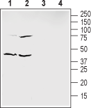
 Western blot analysis of mouse enriched brain membranes (lanes 1 and 3) and human HT-29 colon adenocarcinoma cells lysate (lanes 2 and 4):1,2. Anti-Ephrin-B1 (extracellular) Antibody (#AER-041), (1:200).
3,4. Anti-Ephrin-B1 (extracellular) Antibody, preincubated with Ephrin-B1 (extracellular) Blocking Peptide (#BLP-ER041).
- Kataoka, H. et al. (2002) J. Cancer Res. Clin. Oncol. 128, 343.
- Tanaka, M. et al. (2007) J. Cell Sci. 120, 2179.
- Pasquale, E.B. (2008) Cell 133, 38.
- Flanagan, J.G. et al. (1998) Ann. Rev. Neurosci. 21, 309.
The erythropoietin-producing hepatocellular (EPH) receptors represent the largest known family of receptor tyrosine kinases and are activated by interactions with cell-surface ligands, termed EFNs. EPH receptors have been classified into two subfamilies, EPHA and EPHB, according to their preference for either glycosylphosphatidylinositol (GPI)-anchored EFN-A ligands or transmembrane EFN-B ligands. Thus far, 14 vertebrate receptors and eight ligands have been identified1. All the ligands share a conserved core sequence of approximately 125 amino acids, including 4 invariant cysteine residues, probably corresponding to a receptor binding domain. This is followed by a membrane anchorage domain, taking the form of a GPI anchor for five of the ligands (ephrin-A1 to -A5) or a transmembrane domain for three of them (ephrin-B1 to -B3) 2.
Ephrin-B1 is signals forward or reverse signaling mechanisms. Ephrin-B1 PDZ-dependent reverse signaling controls axon guidance of the corpus callosum, whereas forward signaling is critical for normal craniofacial development2.
Eph receptors and ephrins are highly expressed in the brain and in the developing nervous system, where they are involved in fundamental developmental processes of the nervous system, including axon guidance, axonfasciculation, neural crest cell migration, acquisition of brain subregional identity,and neuronal cell survival. Ephrins are dramatically expressed in a wide range of regions of the vertebrate embryo including the ectoderm, mesoderm, and endoderm3.
Overexpression of B-type ephrin in cancer cells is reported to correlate with high invasion and high vascularity of tumors and elevated expression of ephrin-B1 is observed in poorly differentiated invasive tumor cells and other tumors with poor clinical prognosis4.
Application key:
Species reactivity key:
Alomone Labs is pleased to offer a highly specific antibody directed against an epitope of human Ephrin-B1. Anti-Ephrin-B1 (extracellular) Antibody (#AER-041) can be used in western blot analysis. The antibody recognizes an extracellular epitope and is thus ideal for detecting the ligand in living cells. It has been designed to recognize Ephrin-B1 from human, rat and mouse samples.

