Overview
- Peptide (C)KAGLPRHSFGRNALE, corresponding to amino acid residues 405 - 419 of rat GABRB2 (Accession P63138). Intracellular, 2nd loop.

GABA(A) β2 Receptor Blocking Peptide (BLP-GA012)
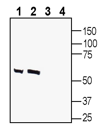
 Western blot analysis of rat brain lysate (lanes 1 and 3) and mouse brain lysate (lanes 2 and 4):1-2. Anti-GABA(A) β2 Receptor Antibody (#AGA-012), (1:200).
3-4. Anti-GABA(A) β2 Receptor Antibody, preincubated with GABA(A) β2 Receptor Blocking Peptide (BLP-GA012). Western blot analysis of human SH-SY5Y neuroblastoma cell line lysate (lanes 1 and 3) and human U-87 MG glioblastoma cell line lysate (lanes 2 and 4):1-2. Anti-GABA(A) β2 Receptor Antibody (#AGA-012), (1:200).
3-4. Anti-GABA(A) β2 Receptor Antibody, preincubated with GABA(A) β2 Receptor Blocking Peptide (BLP-GA012).
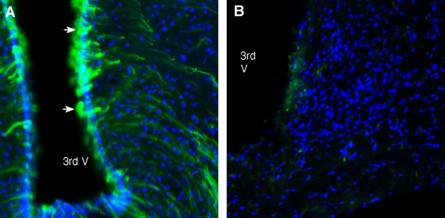
 Expression of GABA(A) Receptor Beta 2 in mouse hypothalamus.Immunohistochemical staining of perfusion-fixed frozen mouse brain sections Anti-GABA(A) β2 Receptor Antibody (#AGA-012), (1:200), followed by goat anti-rabbit-AlexaFluor-488. A. Staining in the periventricular hypothalamus region, showed GABA(A) Receptor Beta 2 immunoreactivity (green) in periventricular glial processes (arrows). B. Pre-incubation of the antibody with GABA(A) β2 Receptor Blocking Peptide (BLP-GA012), suppressed staining. Cell nuclei are stained with DAPI (blue). 3rd V = 3rd ventricle.
 Expression of GABA(A) Receptor Beta 2 in rat hypothalamus.Immunohistochemical staining of perfusion-fixed frozen rat brain sections Anti-GABA(A) β2 Receptor Antibody (#AGA-012), (1:200), followed by goat anti-rabbit-AlexaFluor-488. A. Staining in the periventricular hypothalamus region, showed GABA(A) Receptor Beta 2 immunoreactivity (green) in periventricular glial processes (arrows). B. Pre-incubation of the antibody with GABA(A) β2 Receptor Blocking Peptide (BLP-GA012), suppressed staining. Cell nuclei are stained with DAPI (blue). 3rd V = 3rd ventricle.
- Olsen, R.W. et al. (2018) Neuropharmacology, 136(Pt A): p. 10-22.
- Macdonald, R.L. and R.W. Olsen, et al. (1994) Annu Rev Neurosci, 17: p. 569-602.
- Olsen, R.W. and W. Sieghart. et al. (2008) Pharmacol Rev, 60(3): p. 243-60.
- Olsen, R.W. and W. Sieghart, et al. (2009) Neuropharmacology, 56(1): p. 141-8.
- Barki, M. et al. (2022) Gene. 809, 146021.
GABA receptors have two main subtypes: GABA(A) and GABA(B) receptors based on their amino acid sequence and structure. The GABA(A) receptors are ionotropic receptors while the GABA(B) receptors are metabotropic G-protein coupled receptors1.
To date, 19 GABA(A) receptor subunits have been identified: α 1-6, β 1-3, γ1-3, δ, ε, θ, p and ρ 1-34. GABA(A)receptors are heteropentameric complexes formed by combinations of different GABA(A) receptor subunits 4.
The major isoform of the GABA(A) receptor in the brain is composed of two α1, two β2, and one γ2 subunits that are encoded by the GABRA1, GABRB2, and GABRG2 genes respectively, accounting for 43% of the GABA receptors in the mammalian brain 5. It is then not surprising that GABA(A) Receptor Beta 2, has been associated with several neuropsychiatric disorders such as schizophrenia, bipolar disorder, epilepsy, autism spectrum disorder, Alzheimer’s disease, frontotemporal dementia and depression 5.
Application key:
Species reactivity key:
Anti-GABA(A) β2 Receptor Antibody (#AGA-012) is a highly specific antibody directed against an epitope of the rat protein. The antibody can be used in western blot and immunohistochemistry applications. It has been designed to recognize GABA(A) Receptor Beta 2 from rat, mouse and human samples.

GABA(A) receptors are ion channels opened when GABA or its selective agonist muscimol binds to the receptor. In contrast, when the competitive antagonists bicuculline or gabazine binds to the GABA(A) receptors, the GABA-evoked current is decreased. GABA(A) receptors are blocked with picrotoxin, a non-competitive blocker, that blocks the GABA(A) receptor pore. Benzodiazepines and barbiturates are positive allosteric modulators of GABA(A) receptors and can enhance the currents of these channels2-4.