Overview
- Peptide QGESRRQEPGDFVKQ(C), corresponding to amino acid residues 29-43 of human GABRA3 (Accession P34903). Extracellular, N-terminus.

 Western blot analysis of rat brain membranes:1. Anti-GABA(A) α3 Receptor (extracellular) Antibody (#AGA-003), (1:200).
2. Anti-GABA(A) α3 Receptor (extracellular) Antibody preincubated with GABA(A) α3 Receptor (extracellular) Blocking Peptide (#BLP-GA003).
- Transfected COS-7 cells (1 µg antibody/140 µg lysate) (Zhou, C. et al. (2013) J. Biol. Chem. 288, 21458.).
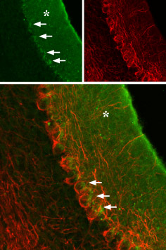
 Expression of GABRA3 in rat cerebellumImmunohistochemical staining of rat cerebellum using Anti-GABA(A) α3 Receptor (extracellular) Antibody (#AGA-003), (green), (1:100). GABRA3 is localized to the molecular layer (asterisk) and a portion of the Purkinje cell body (arrows), which is outlined by axonal staining using mouse anti-neurofilament 200 (red).
 Expression of GABRA3 in rat insulinoma cell lineCell surface detection of GABRA3 in intact living rat RIN-m cells using Anti-GABA(A) α3 Receptor (extracellular) Antibody (#AGA-003), (1:50) followed by goat anti-rabbit-AlexaFluor-550 secondary antibody. Extracellular staining (red) merged with live view of the cells.
- Mihic, S.J. and Harris, R.A. (1997) Alcohol Health Res. World 21, 127.
- Whiting, P.J. (1999) Neurochem. Int. 34, 387.
- Fuchs, K. and Celepirovic, N. (2002) J. Neurochem. 82, 1512.
The neurotransmitter GABA (γ-aminobutiric acid) inhibits the activity of signal-receiving neurons by interacting with the GABAA receptor on these cells.1 Binding of GABA to its GABAA receptor results in conformational changes that open a Cl- channel, producing an increase in membrane conductance that results in inhibition of neural activity.2
There are two major types of GABA receptors: the ionotropic GABAA receptors (GABAAR) and the metabotropic GABAB receptors (GABABR). GABAARs belong to the ligand-gated ion channel superfamily.1,2
GABAARs are heteropentamers, in which all five subunits contribute to formation of the pore. Eight subunit isoforms have been cloned: α, β, γ, δ, ε, π, θ, and ρ.1 Six α subunits isoforms (α1-α6) have been shown to exist in mammals.
In most cases, the native GABAA receptors consist of 2α, 2β, and 1γ subunits.
The α3-subunit is highly expressed during development (along with α2 and α5) and then declines in adulthood, where the α1-subunit becomes predominant. The failure to complete this switch could be a major predispositional factor in the development of temporal lobe epilepsy.3
Application key:
Species reactivity key:
Alomone Labs is pleased to offer a highly specific antibody directed against an epitope of human GABA(A) α3 receptor. Anti-GABA(A) α3 Receptor (extracellular) Antibody (#AGA-003) can be used in western blot, immunoprecipitation, immunocytochemistry, and immunohistochemistry applications. The antibody recognizes an extracellular epitope and is highly suited to detect GABRA3 in living cells. It has been designed to recognize GABRA3 from rat, mouse, and human samples.

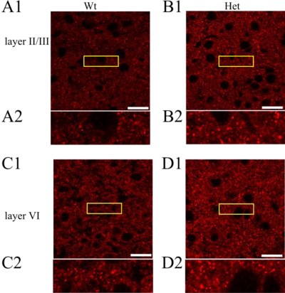
GABA(A) α3 Receptor expression increases in Heterozygous GABA(A) α1 Receptor knockout.Immunohistochemical staining of cortices from wild type (A and C) and Het α1 KO (B and D) mice in layers II/III (A and B) and VI (C and D) of the somatosensory cortex (white scale bar, 20 μm, n > 6) using Anti-GABA(A) α3 Receptor (extracellular) Antibody (#AGA-003). The yellow boxed areas are displayed on a magnified scale below each image (A2–D2).Adapted from Zhou, C. et al. (2013) with permission of the American Society for Biochemistry and Molecular Biology.
Applications
Citations
- Immunocytochemistry staining of rat cultured oligodendrocyte progenitor cells. Tested in cells treated with GABA(A)α3 siRNA.
Ordaz, R.P. et al. (2021) Mol. Pharmacol. 99, 133.
- Mouse brain lysates (1:500).
Seo, S. and Leitch, B. (2014) Epilepsia doi: 10.1111/epi.12500. - Guinea pig and rat medulla.
Inoue, M. et al. (2013) Neuroscience 253, 245. - Mouse brain lysates (1:500).
Zhou, C. et al. (2013) J. Biol. Chem. 288, 21458.
- Rat oligodendrocytes (1:250).
Arellano, R.O. et al. (2016) Mol. Pharmacol. 89, 63.
- Transfected COS-7 cells (1 µg antibody/140 µg lysate).
Zhou, C. et al. (2013) J. Biol. Chem. 288, 21458.
- Rat brain sections (1:100).
Arellano, R.O. et al. (2016) Mol. Pharmacol. 89, 63. - Mouse brain sections (1:200).
Seo, S. and Leitch, B. (2014) Epilepsia doi: 10.1111/epi.12500. - Mouse brain sections (1:500).
Zhou, C. et al. (2013) J. Biol. Chem. 288, 21458. - Rat brain sections (1:250).
Park, H.J. et al. (2013) J. Nucl. Med. 54, 1263.
- Caspary, D.M. et al. (2013) Neurobiol. Aging 34, 1486.

