Overview
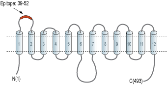
- Peptide (C)KDFLNYTLEERLED, corresponding to amino acid residues 39-52 of rat Glucose Transporter 3 (Accession Q07647). 1st extracellular loop.

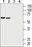
 Western blot analysis of rat brain membranes (lanes 1 and 3) and mouse brain membranes (lanes 2 and 4):1, 2. Anti-GLUT3 (extracellular) Antibody (#AGT-023), (1:400).
3, 4. Anti-GLUT3 (extracellular) Antibody, preincubated with GLUT3 (extracellular) Blocking Peptide (#BLP-GT023).
 Expression of Glucose transporter 3 in mouse hippocampus and cerebellumImmunohistochemical staining of perfusion-fixed frozen mouse brain sections with Anti-GLUT3 (extracellular) Antibody (#AGT-023), (1:200), followed by goat anti-rabbit-AlexaFluor-488. A. GLUT3 staining (green) in mouse hippocampal dentate gyrus, is detected in interneurons (arrows pointing up) in the Hilus and granule layer (G) (arrow pointing down). B. Staining in mouse cerebellum, is seen in purkinje cells (vertical arrows) and dendrites (horizontal arrows) in the molecular layer (Mol). Cell nuclei were stained with DAPI (blue).
- Lacombe, V.A. (2014) ISRN Vet. Sci. 2014, 409547.
- Simpson, I.A. et al. (2008) Am J. Physiol. 295, E242.
- Zheng, C. et al. (2016) Oncol. Lett. 12, 125.
The GLUT protein family is encoded by the SLC2 genes and are a member of the major facilitator superfamily of membrane transporters. GLUTs are recognized as key regulators of whole-body glucose homeostasis, and are responsible for the uptake of carbohydrate-derived glucose by cells. There are 14 GLUT isoforms divided into 3 different group classes based on sequence similarity and structural and functional characteristics1,2.
GLUT3 is a low-Km protein responsible for glucose uptake into neurons. It is predominantly expressed in neurons and is commonly called “neuronal glucose transporter". The protein contains 12 membrane-spanning domains, an N-linked glycosylation site in loop 1 responsible for high-affinity transport of glucose, and intracellular NH2- and COOH- termini. In addition, there are several conserved residues and motifs designated “sugar transporter signatures”1,2.
GLUT3 levels are high in some malignant glioma cells compared to normal glial cells and therefore may be used as a therapeutic target in some cancer treatment3.
Application key:
Species reactivity key:
Anti-GLUT3 (extracellular) Antibody (#AGT-023) is a highly specific antibody directed against an epitope of the rat Glucose Transporter 3 protein. The antibody can be used in western blot, immunohistochemistry, and live cell flow cytometry applications. The antibody has been designed to recognize GLUT3 from rat and mouse samples. It will not recognize GLUT3 from human samples.

