Overview
- Peptide (C)EEFYNQTWNHRYGES, corresponding to amino acid residues 41-55 of rat GLUT1 (Accession P11167). 1st extracellular loop.

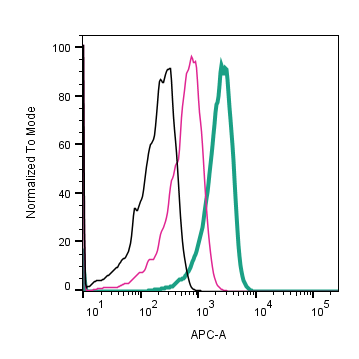
 Cell surface detection of GLUT1 by direct flow cytometry in live intact human Jurkat T-cell leukemia cells:___ Cells.
___ Cells + Rabbit IgG Isotype control-APC (#RIC-001-APC).
___ Cells + Anti-GLUT1 (extracellular)-APC Antibody (#AGT-041-APC), (5 µg). Cell surface detection of GLUT1 by direct flow cytometry in live intact mouse TK-1 T-cell lymphoma cells:___ Cells.
___ Cells + Rabbit IgG Isotype control-APC (#RIC-001-APC).
___ Cells + Anti-GLUT1 (extracellular)-APC Antibody (#AGT-041-APC), (5 µg). Cell surface detection of GLUT1 by direct flow cytometry in live intact mouse J774 macrophage cells:___ Cells.
___ Cells + Rabbit IgG Isotype control-APC (#RIC-001-APC).
___ Cells + Anti-GLUT1 (extracellular)-APC Antibody (#AGT-041-APC), (5 µg).
- Olson, A.L. et al. (1996) Annu. Rev. Nutr 16, 235.
- Pardridge, W.M. et al. (1990) J. Biol. Chem. 265, 18035.
- Klepper, J. et al. (1999) Neurochem. Res. 24, 587.
- Leen, W.G. et al. (2014) J. Neurol. 261, 589.
- Amann, T. et al. (2011) Mol. Membr. Biol. 28, 182.
Glucose transporter 1 (GLUT1) belongs to the major facilitator superfamily (MFS). It is encoded by SLC2A1, and mediates basal-level cellular uptake of glucose into many tissues. GLUT1 contains 12 membrane-spanning domains with both the amino and carboxyl termini oriented intracellularly. In addition, a single extracellular N-linked glycosylation site is present1.
GLUT1 is widely expressed, but it is most abundant in fibroblasts, erythrocytes, and endothelial cells with low levels of expression in muscle, liver, and adipose tissue2.
Inactivating mutations of GLUT1, resulting in compromised transport activities for glucose, are associated with diseases as a result of lack of energy supply to the brain3. GLUT1 deficiency syndrome (also known as De Vivo syndrome) is characterized by a spectrum of symptoms including early-onset seizures, microcephaly and retarded development4. In addition, elevated expression levels of GLUT1 have been observed in several cancer types, identifying GLUT1 as an important prognostic indicator for tumorigenesis5.
Application key:
Species reactivity key:
Anti-GLUT1 (extracellular) Antibody (#AGT-041) is a highly specific antibody directed against an extracellular epitope of the rat protein. The antibody can be used in western blot and indirect live cell flow cytometry. It has been designed to recognize GLUT1 from mouse, rat and human samples.
Anti-GLUT1 (extracellular)-APC Antibody (#AGT-041-APC) is directly conjugated to Allophycocyanin (APC) fluorophore. This conjugated antibody has been developed to be used in immunofluorescent applications such as direct flow cytometry and live cell imaging.
