Overview
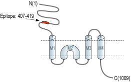
- Peptide (C)KDMRKLATWDSEK, corresponding to amino acid residues 407-419 of rat GluD1 (Accession Q62640). Extracellular, N-terminus.

 Western blot analysis of human CCF-STTG1 astrocytoma cell lysate (lanes 1 and 4), mouse brain lysate (lanes 2 and 5) and rat brain lysate (lines 3 and 6):1-3. Anti-GRID1 (extracellular) Antibody (#AGC-038), (1:200).
4-6. Anti-GRID1 (extracellular) Antibody, preincubated with GRID1 (extracellular) Blocking Peptide (#BLP-GC038).
 Expression of Glutamate receptor δ1 in rat cortex and medial septumImmunohistochemical staining of perfusion-fixed frozen rat brain sections using Anti-GRID1 (extracellular) Antibody (#AGC-038), (1:400). A. Staining in cortex. B. Staining in medial septum. In both regions, GluD1 expression (red) is detected in neurons (arrows). DAPI is used as the counterstain (blue).
 Expression of Glutamate receptor δ1 in rat PC12 cellsCell surface detection of GluD1 in live intact rat PC12 pheochromocytoma cells. A. Extracellular staining of cells with Anti-GRID1 (extracellular) Antibody (#AGC-038), (1:50), followed by goat anti-rabbit-AlexaFluor-594 secondary antibody (red). B. Live view of the cells. C. Merge of A and B.
- Schmid, S.M. et al. (2009) Proc. Natl. Acad. Sci. U.S.A. 106, 10320.
- Traynelis, S.F. et al. (2010) Pharmacol. Rev. 62, 405.
- Lomeli, H. et al. (1993) FEBS Lett. 315, 318.
- Safieddine, S. and Wenthold, R.J. (1997) J. Neurosci. 17, 7523.
- Gao, J. et al. (2007) Mol. Cell Biol. 27, 4500.
- Treutlein, J. et al. (2009) Schizophr. Res. 111, 123.
- Smith, M. et al. (2009) Ann N Y Acad Sci 1151, 102.
- Balciuniene, J. et al. (2007) Am. J. Hum. Genet. 80, 938.
Excitatory neurotransmission in the vertebrate central nervous system is mainly mediated by ionotropic glutamate receptors (iGluRs). Molecular cloning identified 18 mammalian iGluR subunits, of which only 16 sort into the traditional pharmacological subfamilies of AMPA, kainate (KA), and N-methyl-D-aspartate (NMDA) receptors. The 2 remaining subunits were termed “orphan” receptors, “glutamate-like” receptors, “nonionotropic” receptors, or, most commonly, delta receptors1.
Ionotropic glutamate receptors are integral membrane proteins composed of four large subunits that form a central ion channel pore. Sequence similarity among all known glutamate receptor subunits, including the δ receptors, suggests they share a similar architecture. Glutamate receptor subunits are modular structures that contain four discrete semiautonomous domains: the extracellular amino-terminal domain (ATD), the extracellular ligand-binding domain (LBD), the transmembrane domain (TMD), and an intracellular carboxyl-terminal domain (CTD)2.
The delta family of ionotropic glutamate receptors (iGluRs) consists of the glutamate δ1 (GluD1) and glutamate δ2 (GluD2) receptors3. GluD1 is highly expressed in the inner hair cells of the organ of Corti4, diffusely expressed throughout the forebrain during development with high levels in the hippocampus during adulthood3. Deletion of GluD1 leads to a deficit in high frequency hearing in mice5. Genetic association studies have established the GRID1 gene, which codes for GluD1, is a strong candidate gene for schizophrenia, bipolar disorder, and major depressive disorder6. Copy number variation studies have also implicated GRID1 in autism spectrum disorder (ASD)7. In addition, GRID1 gene is localized to the 10q22–q23 genomic region which is a site for recurrent deletions associated with cognitive and behavioral abnormalities8.
Application key:
Species reactivity key:
Alomone Labs is pleased to offer a highly specific antibody directed against an extracellular epitope of the rat Glutamate receptor δ1. Anti-GRID1 (extracellular) Antibody (#AGC-038) can be used in western blot, immunohistochemistry, and live cell imaging applications. It has been designed to recognize GluD1 from rat, mouse, and human samples.
Applications
Citations
- Mouse brain lysate (1:1000).
Suryavanshi, P.S. et al. (2016) Mol. Pharmacol. 90, 96.
