Overview
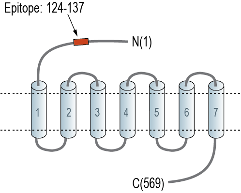
- Peptide (C)KDLQVQVRKYGEQK, corresponding to amino acid residues 124 - 137 of mouse GPR108 (Accession Q91WD0). Extracellular, N-term.

GPR108 (extracellular) Blocking Peptide (BLP-GR088)
 Western blot analysis of rat brain lysate (lanes 1 and 3) and mouse brain lysate (lanes 2 and 4):1-2. Anti-GPR108 (extracellular) Antibody (#AGR-088), (1:200).
3-4. Anti-GPR108 (extracellular) Antibody, preincubated with GPR108 (extracellular) Blocking Peptide (BLP-GR088). Western blot analysis of mouse TK-1 T-cell lymphoma cell line lysate (lanes 1 and 4), human Jurkat T-cell leukemia cell line lysate (lanes 2 and 5) and mouse BV-2 microglia cell line lysate (lanes 3 and 6):1-3. Anti-GPR108 (extracellular) Antibody (#AGR-088), (1:200).
4-6. Anti-GPR108 (extracellular) Antibody, preincubated with GPR108 (extracellular) Blocking Peptide (BLP-GR088).
 Expression of GPR108 in mouse red nucleus.Immunohistochemical staining of perfusion-fixed frozen mouse brain sections with Anti-GPR108 (extracellular) Antibody (#AGR-088), (1:200), followed by goat anti-rabbit-AlexaFluor-488. A. GPR108 immunoreactivity (green) appears in neurons (arrows). B. Pre-incubation of the antibody with GPR108 (extracellular) Blocking Peptide (BLP-GR088), suppressed staining. Cell nuclei are stained with DAPI (blue).
- Fredriksson, R., et al. (2003) Mol Pharmacol, 63(6): p. 1256-72.
- Watkins, L.R. and C. Orlandi. (2020) Genes (Basel), 11(6).
- Stadel, J.M., S. Wilson, and D.J. Bergsma. (1997) Trends Pharmacol Sci, 18(11): p. 430-7.
- Marchese, A., et al. (1999) Trends Pharmacol Sci, 20(9): p. 370-5.
- Edgar, A.J. (2007) DNA Seq, 18(3): p. 235-41.
- Halsey, T.A., et al. (2007) Genome Biol, 8(6): p. R104.
- Dong, D., et al. (2018) PLoS One, 13(10): p. e0205303.
- Dudek, A.M. et al. (2020) Mol. Ther. 28, 367.
G protein coupled receptors (GPCR or GPR) comprise a large superfamily of receptors characterized by a seven transmembrane domain structure and an ability to activate intracellular transducer G proteins. Over 800 GPCRs in five main families have been identified in eukaryotes on the basis of genomic sequence analysis1,2. These receptors can be activated by hormones, neurotransmitters, odorants, light, or pheromones. Whereas the endogenous agonist is remains unclear or in dispute2.
GPCRs mediated the signaling of extracellular stimuli to intracellular responses via activation of heterotrimeric G proteins and their subsequent interaction with effector proteins. GPCRs have been implicated in a number of physiological functions including development of anxiety, behavior, homeostasis, cognition, appetite, and drug addiction3.
The GPCR family contains a diverse group of cell-surface mediators of signal transduction, its size exceeding other cell-surface protein receptors including the tyrosine kinase receptors, guanylyl cyclase receptors and ligand-gated ion channels4.
GPR108 (LUSTR2), has a large extracellular domain of an amino-terminal hydrophobic signal peptide sequence, and a carboxy-terminal seven-transmembrane domain5. Previous studies showed that GPR108 is linked to nuclear factor κB (NF-κB) signaling6 and regulates and inflammatory immune response and cancer signaling7.
Recently, GPR108 has been identified as a highly conserved cellular entry factor for Adeno-associated virus (AAV), a widely used and clinically approved gene therapy vector8.
Application key:
Species reactivity key:
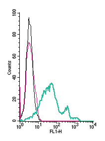
Anti-GPR108 (extracellular) Antibody (#AGR-088) is a highly specific antibody directed against an extracellular epitope of the mouse protein. The antibody can be used in western blot, immunohistochemistry and flow cytometry applications. It has been designed to recognize GPR108 from mouse, rat and human samples.

