Overview
- Peptide (C)RHTGRLHLRYGKND, corresponding to amino acid residues 86-109 of rat GPR56 (Accession Q8K3V3). Extracellular, N-terminus.
Western blot analysis (unlabeled antibody, #AGR-047), and direct flow cytometry (labeled antibody).
- Mouse BV-2 microglia cells, human HL-60 promyelocytic leukemia cells (2.5-5 µg).
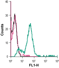
 Cell surface detection of GPR56 in live intact mouse BV-2 microglia cells:___ Cells.
___ Cells + rabbit IgG isotype control-FITC.
___ Cells + Anti-GPR56 (extracellular)-FITC Antibody (#AGR-047-F), (2.5 µg). Cell surface detection of GPR56 in live intact human HL-60 promyelocytic leukemia cells:___ Cells.
___ Cells + rabbit IgG isotype control-FITC.
___ Cells + Anti-GPR56 (extracellular)-FITC Antibody (#AGR-047-F), (5 µg).
GPR56 (ADGRG1) is a member of the adhesion G-protein coupled receptor (GPCR) family, responsible for the regulation of cell adhesion, migration, polarity, and guidance. GPR56 levels are increased in many cancer types and seems to be relevant for tumor cell migration and invasion1,2. GPR56 structure is characterized by a long extracellular domain that is involved in cell-to-cell and cell-to-extracellular matrix interactions. The receptor contains a GPS motif, responsible for cleaving the protein. Seven different glycosylation sites along its N-terminal fragment are present1-3.
Expression of Gpr56 mRNA can be found in neuronal progenitor cells of the cerebral cortical ventricular and subventricular zones during periods of neurogenesis. Studies show that GPR56 protein knockdown promotes mesenchymal differentiation of glioma stem-like initiating cells, accompanied by increased radioresistance in vitro and in vivo1-3.
Mutations in GPR56 may cause bilateral frontoparietal polymicrogyria, a cobblestone-like cortical malformation, characterized by overmigrating neurons and the formation of neuronal ectopias on the surface of the brain2,3.
