Overview
- Peptide (C)RRYGAESQNPTVK, corresponding to amino acid residues 59-71 of mouse GPR83 (Accession P30731). Extracellular, N-terminus.

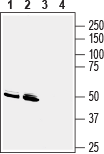
 Western blot analysis of mouse brain membranes (lanes 1 and 3) and rat brain membranes (lanes 2 and 4):1, 2. Anti-GPR83 (extracellular) Antibody (#AGR-053), (1:400).
3, 4. Anti-GPR83 (extracellular) Antibody, preincubated with GPR83 (extracellular) Blocking Peptide (#BLP-GR053). Western blot analysis of human Jurkat T-cell leukemia cell line lysate:1. Anti-GPR83 (extracellular) Antibody (#AGR-053), (1:400).
2. Anti-GPR83 (extracellular) Antibody, preincubated with GPR83 (extracellular) Blocking Peptide (#BLP-GR053).
 Expression of GPR83 in mouse hippocampus and cortexImmunohistochemical staining of perfusion-fixed frozen mouse brain sections with Anti-GPR83 (extracellular) Antibody (#AGR-053), (1:3000), followed by goat anti-rabbit-AlexaFluor-488. A. GPR83 staining (green) in hippocampal dentate gyrus is detected in hilar interneurons (vertical arrows) and around cells of the granule layer (horizontal arrows). H = hilus of dentate gyrus. G = granule layer. B. GPR83 immunoreactivity (green) in parietal cortex is observed in neurons of the pyramidal layer (vertical arrows) and in their apical dendrites (horizontal arrows). Cell nuclei are stained with DAPI (blue).
- Gomes, I. et al. (2016) Sci. Signal. 9, ra43.
- Cherezov, V. et al. (2007) Science 318, 1258.
- Khan, M.Z. and He, L. (2017) Psychopharmacology 24, 1181.
- Sugimoto, N. et al. (2006) Int. Immunol. 18, 1197.
GPR83 (JP05, GIR, GPR72) is a G-protein coupled receptor (GPCR) highly expressed in the brain, spleen and thymus, suggesting that this receptor may be targeted to treat neurological and immune disorders1.
GPCR is a family of receptors responsible for initiation of a myriad of intracellular signaling cascades, these receptors are characterized by an extracellular N-terminus, intracellular C-terminal tail, and seven membrane-spanning α-helices (TM-1 to TM-7)2.
In the brain GPR83 is expressed in the hippocampus, amygdala, prefrontal cortex, and in various hypothalamic nuclei where it may play a significant role in learning memory, reward, emotional behaviors, and stress regulation3. GPR83 is also expressed in regulatory T cells (Tregs) and it could potentially be used as a specific marker used to identify natural Tregs4.
GPR83 was considered as an "orphan" GPCR. Recently, PEN, a neuropeptide was identified as its endogenous ligand1.
Application key:
Species reactivity key:
Anti-GPR83 (extracellular) Antibody (#AGR-053) is a highly specific antibody directed against an epitope of the mouse protein. The antibody can be used in western blot and indirect live cell flow cytometry applications. It has been designed to recognize GPR83 from rat, mouse, and human samples.

