Overview
- Peptide (C)HSRQYVSGLHLNRE, corresponding to amino acids 396-409 of rat HRH1 (Accession P31390). 3rd intracellular loop.

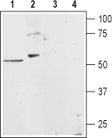
 Western blot analysis of rat heart membranes (lanes 1 and 3) and rat basophilic leukemia (RBL) cell lysates (lanes 2 and 4):1,2. Anti-Histamine H1 Receptor (HRH1) Antibody (#AHR-001), (1:200).
3,4. Anti-Histamine H1 Receptor (HRH1) Antibody, preincubated with Histamine H1 Receptor/HRH1 Blocking Peptide (#BLP-HR001). Western blot analysis of mouse brain membranes:1. Anti-Histamine H1 Receptor (HRH1) Antibody (#AHR-001), (1:400).
2. Anti-Histamine H1 Receptor (HRH1) Antibody, preincubated with Histamine H1 Receptor/HRH1 Blocking Peptide (#BLP-HR001).
 Expression of Histamine H1 receptor in mouse brainImmunohistochemical staining of mouse ventromedial hypothalamus (VMH) using Anti-Histamine H1 Receptor (HRH1) Antibody (#AHR-001). A. H1R (green fluorescence) appears in the outline of the VMH nucleus (arrows). B. DAPI (blue) counterstain labels all cell nuclei including VMH.
- Mouse isolated microglia (Apolloni, S. et al. (2017) Front. Immunol. 8, 1689.).
- Mouse P815 mast cells (Dong, H. et al. (2019) Front. Cell. Neurosci. 13, 191.).
- Hill, S.J. et al. (1997) Pharmacol. Rev. 49, 253.
- Thurmund, R.L. et al. (2008) Nat. Rev. Drug. Discov. 7, 41.
- Haas, H.L. et al. (2008) Physiol. Rev. 88, 1183.
Histamine (2-[4-imidazole]ethylamine) is a low-molecular-weight amine synthesized from L-histidine. It is produced by various cells throughout the body, including central nervous system neurons, gastric mucosa parietal cells, mast cells, basophils and lymphocytes. Histamine is a major biological mediator whose functions include, among many others, regulation of vascular smooth muscle, immune regulation, regulation of sleep-wake cycles and regulation of gastric acid secretion.1
The biological effects of histamine are mediated through four receptors (H1-H4 receptors) all of which belong to the 7-transmembrane domain, G protein-coupled receptor (GPCR) superfamily.
H1 receptors couple to Gq/G11 proteins leading to phospholipase C activation, inositol phosphate production and calcium mobilization.1
H1 receptors are expressed in many cell types including endothelial and smooth muscle cells where they mediate vasodilatation and increased vascular permeability effects. H1R is widely distributed through the brain where it mediates sleep-wake cycles and regulates cognitive functions. The receptor is expressed in several cell populations involved in the immune response, including T-cells and B-cells, monocytes and eosinophiles.2,3
H1 anti-histamine compounds have been available for almost thirty years and are among the most commonly used medication for conditions such as allergic inflammation. In addition, H1 anti-histamines are also prescribed for the treatment of insomnia and analgesia.2,3
Application key:
Species reactivity key:
Anti-Histamine H1 Receptor (HRH1) Antibody (#AHR-001) is a highly specific antibody directed against an epitope of rat H1R. The antibody can be used in western blot, immunocytochemistry, indirect flow cytometry, and immunohistochemstry applications. It has been designed to recognize H1R from rat, mouse, and human samples.

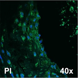
Expression of Histamine H1 Receptor in mouse aortic root.Immunohistochemical staining of mouse aortic root sections using Anti-Histamine H1 Receptor (HRH1) Antibody (#AHR-001). H1R staining is shown in green.Adapted from Raveendran, V.V. et al. (2014) PLoS ONE 9, e10265. with permission of PLos.
Applications
Citations
- Rat primary astrocyte lysate (1:200).
Xu, J. et al. (2018) J. Neuroinflammation 15, 41. - Mouse brain and spinal cord lysate.
Apolloni, S. et al. (2017) Front. Immunol. 8, 1689. - Rat brain lysate (1:400).
Cilz, N.I. and Lei, S. (2017) Hippocampus 27, 613.
- Mouse aortic root sections.
Raveendran, V.V. et al. (2014) PLoS ONE 9, e10265.
- Rat primary astrocytes.
Xu, J. et al. (2018) J. Neuroinflammation 15, 41. - Mouse isolated microglia.
Apolloni, S. et al. (2017) Front. Immunol. 8, 1689.
- Mouse P815 mast cells.
Dong, H. et al. (2019) Front. Cell. Neurosci. 13, 191.
