Overview
- Peptide (C)RTRLRLDGGREAGPE, corresponding to amino acids 228-242 of rat HRH3 (Accession Q9QYN8). 3rd intracellular loop.

 Western blot analysis of rat (lanes 1 and 3) and mouse (lanes 2 and 4) brain membranes:1,2. Anti-Histamine H3 Receptor (HRH3) Antibody (#AHR-003), (1:200).
3,4. Anti-Histamine H3 Receptor (HRH3) Antibody, preincubated with Histamine H3 Receptor/HRH3 Blocking Peptide (#BLP-HR003).
 Expression of Histamine H3 receptor in rat cerebellumImmunohistochemical staining of rat brain frozen sections with Anti-Histamine H3 Receptor (HRH3) Antibody (#AHR-003), (1:100), (green). A. H3R is particularly expressed in dendrites of Purkinje cells (arrows). B. Staining with mouse anti-parvalbumin (red) detected Purkinje cells and interneurons in the molecular layer. C. Merge of the two images demonstrates that the staining was restricted to dendrites of Purkinje cells. Cell nuclei were labeled with DAPI (blue) as the counterstain.
- Rat astrocytes (1:100) (Mele, T. and Juric, D.M. (2013) Eur. J. Pharmacol. 720, 198.).
- Hill, S.J. et al. (1997) Pharmacol. Rev. 49, 253.
- Leurs, R. et al. (2005) Nat. Rev. Drug Discov. 4, 107.
- Esbenshade, T.A. et al. (2006) Mol. Interv. 6, 77.
Histamine (2-[4-imidazole]ethylamine) is a low-molecular-weight amine synthesized from L-histidine. It is produced by various cells throughout the body, including central nervous system neurons, gastric mucosa parietal cells, mast cells, basophils and lymphocytes. Histamine is a major biological mediator whose functions include, among many others, regulation of vascular smooth muscle, immune regulation, regulation of sleep-wake cycles and regulation of gastric acid secretion.1
The biological effects of histamine are mediated through four receptors (H1-H4 receptors) all of which belong to the 7-transmembrane domain, G protein-coupled receptor (GPCR) superfamily.
H3 receptor couples to Gi/G0 proteins and receptor activation leads to inhibition of adenylate cyclase, activation of the mitogen-activated protein kinase (MAPK) cascade and inhibition of the Na+/H+ exchanger.1,2
H3 receptors are expressed primarily in the central nervous system (CNS) where they are located in presynaptic membranes of histaminergic neurons, where they negatively regulate the synthesis and release of histamine. In addition, H3 receptors are also located on nonhistaminergic neurons, where they regulate the release of other amines such as dopamine, serotonin, and norepinephrine.2,3
Based on these studies, a central role for H3 receptors has been proposed in disorders involving cognition such as attention deficit and hyperactivity disorder (ADHD), Alzheimer disease and Schizophrenia, as well as sleep and energy homeostasis (i.e. obesity) disorders.2,3
Application key:
Species reactivity key:
Alomone Labs is pleased to offer a highly specific antibody directed against an epitope of the rat histamine H3 receptor. Anti–Histamine H3 Receptor (HRH3) Antibody (#AHR-003) can be used in western blot, immunocytochemistry and immunohistochemistry applications. It has been designed to recognize H3R from rat, mouse and human samples.

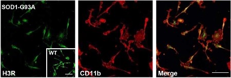
Expression of Histamine H3 Receptor in mouse primary microglia.Immunocytochemical staining of mouse primary microglia using Anti-Histamine H3 Receptor (HRH3) Antibody (#AHR-003). H3R expression (green) partially co-localizes with CD11b (red).Adapted from Apolloni, S. et al. (2017) Front. Immunol. 8, 1689. with permission of Frontiers.
Applications
Citations
- Mouse isolated microglia.
Apolloni, S. et al. (2017) Front. Immunol. 8, 1689.
- Mouse primary microglia.
Apolloni, S. et al. (2017) Front. Immunol. 8, 1689. - Rat astrocytes (1:100).
Mele, T. and Juric, D.M. (2013) Eur. J. Pharmacol. 720, 198.
