Overview
- Peptide CGQPKEGKNHSQG, corresponding to amino acid residues 146 - 158 of human adenosine A2A receptor (Accession P29274). 2nd extracellular loop.

 Western blot analysis of human HL-60 promyelocytic leukemia cell line lysate (lanes 1 and 3) and human Jurkat T-cell leukemia cell line lysate (lanes 2 and 4):1,2. Anti-Human Adenosine A2A Receptor (extracellular) Antibody (#AAR-007), (1:200).
3,4. Anti-Human Adenosine A2A Receptor (extracellular) Antibody, preincubated with Human Adenosine A2A Receptor (extracellular) Blocking Peptide (#BLP-AR007).
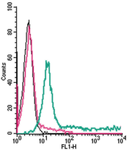
 Cell surface detection of ADORA2A in live intact human THP-1 monocytic leukemia cells:___ Cells.
___ Cells + goat-anti-rabbit-FITC.
___ Cells + Anti-Human Adenosine A2A Receptor (extracellular) Antibody (#AAR-007), (2.5 µg) + goat-anti-rabbit-FITC. Cell surface detection of ADORA2A in live intact human Jurkat T-cell leukemia cells:___ Cells.
___ Cells + goat-anti-rabbit-FITC.
___ Cells + Anti-Human Adenosine A2A Receptor (extracellular) Antibody (#AAR-007), (2.5 µg) + goat-anti-rabbit-FITC.
- Okusa, M.D. (2002) Am. J. Physiol. 282, F10.
- Fredholm, B.B. et al. (2001) Pharmacol. Rev. 53, 527.
- Nakata, H. (1989) J. Biol. Chem. 264, 16545.
- Cunha, R.A. (2001) Neurochem. Int. 38, 107.
- Fredholm, B.B. et al. (2000) Naunyn-Schmiedebergs Arch. Pharmacol. 362, 364.
- Canals, M. et al. (2005) J. Neurochem. 92, 337.
- Ohta, A. and Sitkovsky, M. (2001) Nature 414, 916.
- Ohta, A. et al. (2006) Proc. Natl. Acad. Sci. 103, 13132.
Adenosine is an endogenous nucleoside generated locally in tissues under conditions of hypoxia, ischemia, or inflammation. It modulates a variety of physiological functions in many tissues including the brain and heart1,2. Adenosine exerts its actions via four specific adenosine receptors (also named P1 purinergic receptors): Adenosine A1 Receptor (A1AR), Adenosine A2A Receptor (A2AAR), Adenosine A2B Receptor (A2BAR), and Adenosine A3 Receptor (A3AR). All are integral membrane proteins and are members of the G protein-coupled receptor superfamily. They share a common structure of seven putative transmembrane domains, an extracellular -NH2 terminus, cytoplasmic -COOH terminus, and a third intracellular loop important for binding G proteins.1-3 The adenosine receptors can be distinguished on the basis of their differential selectivity for adenosine analogs1-3.
Adenosine receptors control neurotransmitter release through the facilitatory A2AAR and the inhibitory A1AR.4 A2AAR and A1AR are the major adenosine receptor subtypes expressed in the central nervous system (CNS). A2AAR is mainly expressed in the striatum on GABAergic striatopallidal neurons, while A1AR is widely distributed throughout the CNS5,6.
A2AAR was suggested to play a critical role in attenuation of systemic inflammatory responses and prevention of extensive tissue damage.7 It was suggested that extracellular adenosine that accumulates in inflamed and damaged tissue may activate the A2AAR expressed in immune cells leading to termination/inhibition of the immune response.7 It was further suggested that this same mechanism may protect tumors from antitumor T cells through an immunosuppressive signal generated by the activation of A2AAR on T cells by extracellular adenosine produced from hypoxic cancerous tissues8.
Application key:
Species reactivity key:
Anti-Human Adenosine A2A Receptor (extracellular) Antibody (#AAR-007) is a highly specific antibody directed against an epitope of the human protein. The antibody can be used in western blot and live cell flow cytometry applications. It has been designed to recognize A2AR from human samples only. The antibody will not recognize the receptor from mouse or rat samples.
Applications
Citations
- Human:
Dierks, A. et al. (2019) Cell Physiol. Biochem. 53, 606.
