Overview
- GST fusion protein with amino acid residues 1978-2016 of human NaV1.5 (Accession Q14524), (MW: 33 kDa.). Intracellular, C-terminus.

 Western blot analysis of NaV1.5 in NaV1.5 transfected HEK-293 cells:1. Anti-NaV1.5 (SCN5A) (1978-2016) Antibody (#ASC-013), (1:200).
2. Anti-NaV1.5 (SCN5A) (1978-2016) Antibody, preincubated with Nav1.5/SCN5A (1978-2016) Blocking Peptide (#BLP-SC013).
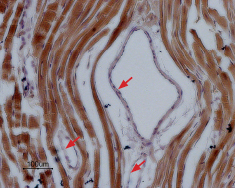
 Expression of NaV1.5 in rat cardiac muscleImmunohistochemical staining of NaV1.5 in rat myocardium paraffin-embedded section using Anti-NaV1.5 (SCN5A) (1978-2016) Antibody (#ASC-013), (1:100). Staining is specific for cardiomyocytes while smooth muscles cells in the artery walls are negative (red arrows). Hematoxilin is used as the counterstain.
- Human atrial myofibroblasts (1:200) (Chatelier, A. et al. (2012) J. Physiol. 590, 4307.).
- THP-1 human premyelomonocytic leukemic cells (Carrithers, M.D. et al. (2007) J. Immunol. 178, 7822.).
- The control antigen is not suitable for this application.
Voltage-gated Na+ channels (NaV) are responsible for myocardial conduction and maintenance of the cardiac rhythm and are essential for the generation of action potentials and cell excitability.1
Dysfunction or disregulation of cardiac sodium channels can cause several disorders, including cardiac arrhythmias.
The majority of Na+ channels in the mammalian heart are Tetrodotoxin (TTX) insensitive NaV1.5.2
The putative structure of NaV1.5 consists of four homologous domain (I-IV), each containing six transmembrane segments (S1-S6). Mutations in the C-terminus of NaV1.5 were described in connection to Long QT syndrome and Brugada syndrome.1-2 Recent data have demonstrated selective expression of NaV1.5 in the mouse central nervous system and implicated a role for NaV1.5 in the physiology of the central nervous system.1
Application key:
Species reactivity key:
Alomone Labs is pleased to offer a highly specific antibody directed against an epitope of human NaV1.5 channel. Anti-NaV1.5 (SCN5A) (1978-2016) Antibody (#ASC-013) can be used in western blot, indirect flow cytometry, immunohistochemistry, and immunocytochemistry applications. It has been designed to recognize NaV1.5 sodium channel from rat, human, and mouse samples.

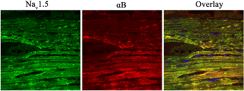
Expression of NaV1.5 in rat heart.Immunohistochemical staining of rat heart sections using Anti-NaV1.5 (SCN5A) (1978-2016) Antibody (#ASC-013). NaV1.5 staining (green), (left panel). αB-Crystallin staining (red), (middle panel). Merged image (right panel) demonstrates that the two proteins co-localize in rat heart.Adapted from Huang, Y. et al. (2016) J. Biol. Chem. 291, 11030. With permission of The American Society for Biochemistry and Molecular Biology.
Applications
Citations
 Multiplex staining of neuronal NaV channels and Ryanodine receptor 2 in mouse cardiomyocytes.Immunocytochemical staining of mouse cardiomyocytes using Anti-SCN1A (NaV1.1) Antibody (#ASC-001), Anti-NaV1.5 (SCN5A) (1978-2016) Antibody (#ASC-013) and Anti-NaV1.6 (SCN8A) Antibody (#ASC-009) antibodies. All three neuronal NaV channels (green) co-localize with Ryanodine receptor 2.
Adapted from Radwanski, P.B. et al. (2015) with permission of the European Society of Cardiology.
- HEK 293 transfected cells.
Beltran-Alvarez, P. et al. (2013) FEBS Lett. 587, 3159. - Human cardiac cells.
Tarradas, A. et al. (2013) PLoS ONE 8, e53220.
- Rat heart sections.
Huang, Y. et al. (2016) J. Biol. Chem. 291, 11030.
- Mouse isolated myocytes.
Radwanski, P.B. et al. (2015) Cardiovasc. Res. 106, 143. - Human atrial myofibroblasts (1:200).
Chatelier, A. et al. (2012) J. Physiol. 590, 4307. - THP-1 human premyelomonocytic leukemic cells.
Carrithers, M.D. et al. (2007) J. Immunol. 178, 7822.
- THP-1 human premyelomonocytic leukemic cells.
Carrithers, M.D. et al. (2007) J. Immunol. 178, 7822.
