Overview
- Peptide CEEIPGRWITRSTPPE corresponding to amino acid residues 188-203 of human p75NTR (Accession P08138). Extracellular, stalk domain.

 Multiplex staining of LINGO-1 and p75NTR in rat hippocampusImmunohistochemical staining of immersion-fixed, free floating rat brain frozen sections using Anti-LINGO-1 (extracellular) Antibody (#ANT-032), (1:200) and Anti-p75 NGF Receptor (extracellular)-ATTO Fluor-550 Antibody (#ANT-007-AO), (1:80). A. LINGO-1 staining (green) in hippocampal CA3 region is detected in neuronal profiles (arrows). B. p75NTR staining in the same section (orange) is also seen in CA3 neurons (arrows). C. Merge of the two images demonstrates colocalization in several neurons (arrows). Cell nuclei are stained with DAPI (blue).
 Expression of p75NTR in p75NTR-transfected 3T3 cellsA. Cell surface detection of p75NTR in intact live p75NTR-transfected 3T3 cells with Anti-p75 NGF Receptor (extracellular)-ATTO Fluor-550 Antibody (#ANT-007-AO), (1:100). B. Live view of the same field as in A.
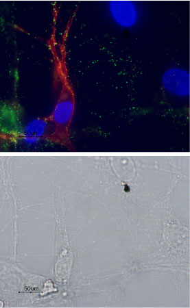
 Multiplex staining of p75NTR and mGluR1 in rat DRGsCell surface detection of p75NTR and mGluR1 in intact live DRG neurons labeled with Anti-mGluR1 (extracellular) Antibody (#AGC-006), (1:100) followed by goat-anti-rabbit-AlexaFluor-488 secondary antibody (green). The cells were then labeled with Anti-p75 NGF Receptor (extracellular)-ATTO Fluor-550 Antibody (#ANT-007-AO), (1:50), (red). Nuclei were visualized using the cell-permeable DNA binding dye Hoechst 33342 (blue). Note that the Anti-p75 NGF Receptor (extracellular)-ATTO Fluor-550 Antibody does not stain all DRG neurons as expected. Colocalization is observed in some nerve fibers of the mGluR1 and p75NTR receptors.
- Hempstead, B.L. (2002) Curr. Opin. Neurobiol. 12, 260.
- Chao, M.V. (2003) Nat. Rev. Neurosci. 4, 299.
- Barker, P.A. (2004) Neuron 42, 529.
- Dechant, G. and Barde, Y.A. (2002) Nat. Neurosci. 5, 1131.
The p75 neurotrophin receptor (p75NTR) is a member of the TNF receptor superfamily. p75NTR, like all the superfamily members is a type I transmembrane protein with tandem cysteine-rich domains in the extracellular portion and a intracellular death domain. p75NTR binds its ligands as a homodimer but can also form heterodimers with other receptors such as TrkA, TrkB, TrkC, Nogo receptor and sortilin. The precise multimeric receptor complex formed between p75NTR and the other receptors will determine the ligand being recognized (see below) and the biological response to its binding.
As it name implies, p75NTR binds all the neurotrophins (NGF, BDNF, NT-3 and NT-4) with similar nM affinities. Co-expression of p75NTR with the Trk receptors enhances the ability to bind and respond to the specific neurotrophin and induces cell survival. On the other hand, it has recently been demonstrated that p75NTR binds with high affinity to the unprocessed form of NGF (proNGF) probably as a complex with sortilin and this leads to cell death by apoptosis. Finally, a multimeric complex of p75NTR and Nogo receptor binds myelin proteins such as Nogo-66, MAG and OmgP resulting in inhibition of axonal growth.
Application key:
Species reactivity key:
Anti-p75 NGF Receptor (extracellular) Antibody (#ANT-007) is a highly specific antibody directed against an extracellular epitope of the human protein. The antibody can be used in western blot, immunoprecipitation, immunohistochemistry, live cell imaging and indirect flow cytometry applications. It has been designed to recognize p75NTR from human, mouse and rat samples.
Anti-p75 NGF Receptor (extracellular)-ATTO Fluor-550 Antibody (#ANT-007-AO) is directly labeled with an ATTO-550 fluorescent dye. ATTO dyes are characterized by strong absorption (high extinction coefficient), high fluorescence quantum yield, and high photo-stability. The ATTO-550 fluorescent label is related to the well known dye Rhodamine 6G and can be used with filters typically used to detect Rhodamine. Anti-p75 NGF Receptor (extracellular)-ATTO Fluor-550 Antibody is especially suited for experiments requiring simultaneous labeling of different markers.
Expression and visualization of p75NTR in live intact dorsal root ganglion (DRG) explants DRG intact explants were incubated with Anti-p75 NGF Receptor (extracellular)-ATTO Fluor-550 Antibody (#ANT-007-AO), (1:50), under conditions that allowed receptor internalization. Retrograde transport of the internalized receptor (orange) was visualized in vesicles moving across the axon. Right side is distal axon while left is explant side. Frames were taken every 2 seconds. Movie is 10 frames per second.
Movie was kindly provided by Dr. Eran Perlsson, the Dept. of Pharmacology and Physiology, Tel-Aviv University.
Applications
Citations
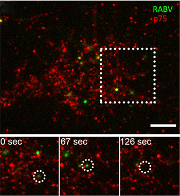
 Co-localization of RABV with p75NTR in DRG neuron tips.Immunocytochemical staining of mouse DRGs using TIRF imaging. RABV-EGFP and Anti-p75 NGF Receptor (extracellular)-ATTO Fluor-550 Antibody (#ANT-007-AO) show that the virus and p75NTR particles shift from the periphery to the center of the growth cone, where they are internalized into the cell. Lower panel show co-localized puncta (left) shifting towards the center of the growth cone (middle) until internalized (right).
Adapted from Gluska, S. et al. (2014) with kind permission from Prof. Eran Perlson, Dpt. Physiology and Pharmacology, Sackler Faculty of Medicine, Tel Aviv University, Israel.
- mouse DRGs.
Gluska, S. et al. (2014) PLoS Pathog. 10, e1004348.
