Overview
- Peptide CEEIPGRWITRSTPPE, corresponding to amino acid residues 188-203 of human p75NTR (Accession P08138). Extracellular, stalk domain.

 Western blot analysis of rat brain membranes:1. Anti-p75 NGF Receptor (extracellular) Antibody (#ANT-007), (1:200).
2. Anti-p75 NGF Receptor (extracellular) Antibody, preincubated with p75 NGF Receptor (extracellular) Blocking Peptide (#BLP-NT007). Western blot analysis of human melanoma cells A875:1. Anti-p75 NGF Receptor (extracellular) Antibody (#ANT-007), (1:200).
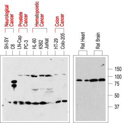
2. Anti-p75 NGF Receptor (extracellular) Antibody, preincubated with p75 NGF Receptor (extracellular) Blocking Peptide (#BLP-NT007). Western blot analysis of normal rat tissue (right) and in human cancer cell lines (left):p75NTR is visualized with Anti-p75 NGF Receptor (extracellular) Antibody (#ANT-007), (1:200). Note that human cancer cell lines from hemapoietic origin show high p75NTR expression, while cell lines from prostate and colon cancer origin show lower levels. Interestingly, p75NTR from rat (right blot and the C6 cell line) and human (left blot) samples run with a different apparent MW, probably due to species-specific differential glycosylation.
 Immunoprecipitation of 3T3/p75NTR transfected cells:1. Cell lysate + protein A beads.
2. Cell lysate + protein A beads + pre-immune rabbit serum.
3. Cell lysate + protein A beads + Anti-p75 NGF Receptor (extracellular) Antibody (#ANT-007).
4. Cell lysate.
Red arrows indicate NGFR p75 while the black arrow shows the IgG heavy chain.
Immunoblot was performed with the Anti-p75 NGF Receptor (extracellular) Antibody.
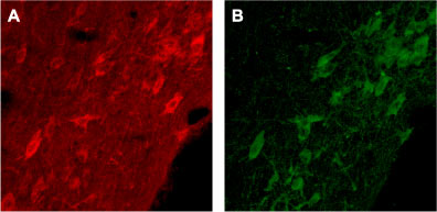
 Expression of p75NTR in rat brainImmunohistochemical staining of rat brain with Anti-p75 NGF Receptor (extracellular) Antibody (#ANT-007). A. Cells in the diagonal band are stained positive for p75NTR. B. Staining of the same section with goat anti-ChAT confirms that p75NTR staining is specific to cholinergic neurons.
 Expression of p75NTR in rat brainImmunohistochemical staining of rat brain with Anti-p75 NGF Receptor (extracellular) Antibody (#ANT-007). A. Cells in the nucleus basalis mangocellularis are stained positive for p75NTR. B. Staining of the same section with goat anti-ChAT confirms that the p75NTR staining is specific to cholinergic neurons.
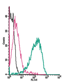
 Cell surface detection of p75NTR by indirect flow cytometry in live intact rat PC12 pheochromocytoma cells:___ Cells.
___ Cells + goat-anti-rabbit-FITC.
___ Cells + Anti-p75 NGF Receptor (extracellular) Antibody (#ANT-007), (2.5μg) + goat-anti-rabbit-FITC. Cell surface detection of p75NTR by indirect flow cytometry in live intact human A-875 Melanoma cells:___ Cells.
___ Cells + goat-anti-rabbit-FITC.
___ Cells + Anti-p75 NGF Receptor (extracellular) Antibody (#ANT-007), (2.5μg) + goat-anti-rabbit-FITC.
- A875 human live melanoma cells lines (1:100-1:200).
- Hempstead, B.L. (2002) Curr. Opin. Neurobiol. 12, 260.
- Chao, M.V. (2003) Nat. Rev. Neurosci. 4, 299.
- Barker, P.A. (2004) Neuron 42, 529.
The p75 neurotrophin receptor (p75NTR) is a member of the TNF receptor superfamily. p75NTR, like all the superfamily members is a type I transmembrane protein with tandem cysteine-rich domains in the extracellular portion and a intracellular death domain. p75NTR binds its ligands as a homodimer but can also form heterodimers with other receptors such as TrkA, TrkB, TrkC, Nogo receptor and sortilin. The precise multimeric receptor complex formed between p75NTR and the other receptors will determine the ligand being recognized (see below) and the biological response to its binding.
As it name implies, p75NTR binds all the neurotrophins (NGF, BDNF, NT-3 and NT-4) with similar nM affinities. Co-expression of p75NTR with the Trk receptors enhances the ability to bind and respond to the specific neurotrophin and induces cell survival. On the other hand, it has recently been demonstrated that p75NTR binds with high affinity to the unprocessed form of NGF (proNGF) probably as a complex with sortilin and this leads to cell death by apoptosis. Finally, a multimeric complex of p75NTR and Nogo receptor binds myelin proteins such as Nogo-66, MAG and OmgP resulting in inhibition of axonal growth.
Application key:
Species reactivity key:
Anti-p75 NGF Receptor (extracellular) Antibody (#ANT-007) is a highly specific antibody directed against an extracellular epitope of the human protein. The antibody can be used in western blot, immunoprecipitation, immunohistochemistry, live cell imaging and indirect flow cytometry applications. It has been designed to recognize p75NTR from human, mouse and rat samples.
Applications
Citations
- Western blot and immunohistochemistry of human A875 melanoma cell line. Tested in A875 cell lines with shRNA knockdown of p75NTR.
Goh, E.T.H. et al. (2018) Cell Chem. Biol. 25, 1485.
- Rat PC12 cells (also used to observe internalization of the receptor).
Escudero, C.A. et al. (2014) J. Cell Sci. 127, 1966.
- Civenni, G. et al. (2011) Cancer Res. 71, 3098.
- D'Onofrio, M. et al. (2011) PLoS One. 6, e20839.
- Wang, Y-J. et al. (2011) J. Neurosci. 31, 2292.
- Chakravarthy, B. et al. (2010) Biochem. Biophys. Res. Com. 401, 458.
- Curchoe, C.L. et al. (2010) PLoS One. 5, e13890.
- Rieske, P. et al. (2009) Exp. Cell Res. 315, 462.
- Ben-Zvi, A. et al. (2007) J. Neurosci. 27, 13000.
- Gil, C. et al. (2007) FEBS Lett. 581, 1851.
- Sandoval, M. et al. (2007) J. Neurochem. 101, 1672.
