Overview
- Peptide (C)DEDLQSKLEAFKTK, corresponding to amino acid residues 40-53 of rat IBA1/AIF1 (Accession P55009). Intracellular.
- Rat spleen, rat testis and mouse spleen (1:200-1:1000).
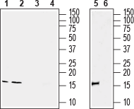
 Western blot analysis of rat spleen (lanes 1 and 3), rat testis (lanes 2 and 4) and mouse spleen (lanes 5 and 6) lysates:1, 2, 5. Anti-IBA1/AIF1 Antibody (1:200), (#ACS-010).
3, 4, 6. Anti-IBA1/AIF1 Antibody, preincubated with IBA1/AIF1 Blocking Peptide (#BLP-CS010).
- Rat brain sections.
Allograft Inflammatory Factor-1 (AIF1) is a 17 kDa. cytoplasmic, calcium and actin binding scaffold protein, mainly expressed in immunocytes. AIF-1 is induced by cytokines such as interferon-γ (IFN-γ).
It is encoded within the HLA class III genomic region by IBA1 gene, and its structure contains seven alpha helixes and three beta sheets. There are three other proteins identical to AIF1: Ionized Ca2+-Binding Adaptor 1, Microglia Response Factor-1, and Daintain1.
The gene is highly expressed in testis, spleen, circulating macrophages and in the brain but weakly expressed in lung and kidney. Among brain cells, the Iba1 gene is strongly and specifically expressed in microglia and its expression is up-regulated following nerve injury, central nervous system ischemia, and several other brain diseases2.
AIF1 influences the immune system at several key points and thus modulates inflammatory diseases. It increases the expression of inflammatory mediators such as cytokines, chemokines, inducible nitric oxide synthase (iNOS) and promotes inflammatory cell proliferation and migration and hence it is highly associated with autoimmune diseases3. In addition, its abundance is positively correlated with metabolic indicators, such as body mass index, triglycerides, and fasting plasma glucose levels. There is also evidence that AIF1 could be a marker for diabetic nephropathy when detected in serum. It is found in activated macrophages in the pancreatic islets, and has been shown to decrease insulin secretion, while simultaneously impairing glucose elimination4.
Application key:
Species reactivity key:

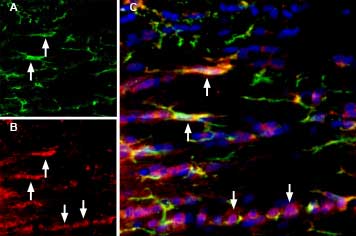
Multiplex staining of IBA1/AIF1 and P2X7 in rat brain.
Immunohistochemical staining of rat corpus callosum (CC) free floating frozen sections using Anti-IBA1/AIF1 Antibody (#ACS-010), (1:1000) and Anti-P2X7 Receptor-ATTO Fluor-550 Antibody (#APR-004-AO) (1:60). A. IBA1/AIF1 immunoreactivity (green) appears in microglia (arrows). B. P2X7 immunostaining (red) appears in microglia (up-pointing arrows) and in other cell types in the corpus callosum (down-pointing arrows). C. Merged image of panels A and B demonstrates partial colocalization of both proteins. Nuclei are demonstrated using DAPI as the counterstain (blue).
