Overview
- Peptide DPKSAAQNSKPRLSFSTK(C), corresponding to residues 8-25 of human KCNK2 (Accession O95069). Intracellular, N-terminus.
- Rat and mouse brain membranes (1:800- 1:4000).
 Western blot analysis of rat brain membranes (lanes 1 and 3) and mouse brain membranes (lanes 2 and 4):1,2. Guinea pig Anti-KCNK2 (TREK-1) Antibody (#APC-047-GP), (1:800).
3,4. Guinea pig Anti-KCNK2 (TREK-1) Antibody, preincubated with KCNK2/TREK-1 Blocking Peptide (#BLP-PC047).
- Rat brain sections (1:400).
KCNK2 (also named TWIK-Related K+ channel, TREK-1 or K2P2.1) is a member of the 2-pore (2P) domain K+ channels family that includes at least 16 members. These channels show little time- or voltage-dependence and are considered to be “leak” or “background” K+ channels, thereby generating background currents which help set the membrane resting potential and cell excitation.
The K2P channels have a signature topology that includes four transmembrane domains and two pore domains with intracellular N- and C- termini.
K2P channels are regulated by diverse physical and chemical stimuli including temperature, changes in intracellular pH, mechanical stretch, inhalation anesthetics, etc. The channels can then be subclassified based in their specific activators. KCNK2 can be integrated to a K2P subfamily that includes K2P4.1 (TRAAK) and K2P10.1 (TREK2) that are activated by intracellular unsaturated fatty acids such as arachidonic acid, lysophosphatidic acid and mechanical stretch. In addition, KCNK2 can also be activated by general anesthetics such as halothane and chloroform and intracellular acidification.
KCNK2 expression in humans is largely restricted to the brain with some expression in ovary and small intestine while KCNK2 expression in rodents is more widespread.
KCNK2 has an important role in mood regulation, as knockout mice show resistance to depression, suggesting that KCNK2 may be a potential target for anti-depressants.
KCNK2 is involved in the fast release of glutamate from astrocytes. It requires the activation of Gαi, dissociation of Gβγ, followed by the opening of the glutamate permeable KCNK2 (TREK-1) K+ channel through its interaction with Gβγ. For this purpose, KCNK2 is localized at the cell surface of the cell body and processes of astrocytes.
Application key:
Species reactivity key:
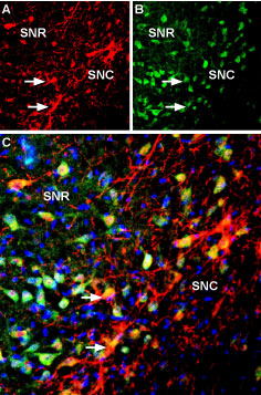
 Multiplex staining of Kir3.2 and KCNK2 in rat substantia nigra.Immunohistochemical staining of immersion-fixed, free floating rat brain frozen sections using rabbit Anti-GIRK2 (Kir3.2) Antibody (#APC-006), (1:400) and Guinea pig Anti-KCNK2 (TREK-1) Antibody (#APC-047-GP), (1:120). A. Kir3.2 staining (red) appears in cells of the substantia nigra pars compacta (SNC, arrows). B. KCNK2 (green) appears in both compacta (SNC) and reticulata (SNR) portions of the substantia nigra. C. Merge of the two images reveals co-localization in some cells (arrows), mainly in the SNC region. Cell nuclei are stained with DAPI (blue).
