Overview
- Peptide (C)EDEKRDAEHRALLTRNGQ, corresponding to amino acid residues 252-269 of human KCNK3 (Accession O14649). Intracellular, C-terminal part.

 Western blot analysis of rat brain (1, 3) and pancreas (2, 4) membranes:1,2. Anti-KCNK3 (TASK-1) Antibody (#APC-024), (1:200).
3,4. Anti-KCNK3 (TASK-1) Antibody, preincubated with KCNK3/TASK-1 Blocking Peptide (#BLP-PC024).
- Transfected HeLa cells (1:1000) (Renigunta, V. et al. (2014) Mol. Biol. Cell 25, 1877.).
- Rat brain sections.
- Human bronchial epithelial cells (Telles, C.J. et al. (2016) Am. J. Physiol. 311, C884.).
- Duprat, F. et al. (1997) EMBO J. 16, 5464.
- Lesage, F. and Lazdunski, M. (2000) Am. J. Physiol. Renal Physiol. 279, F793.
- Bayliss, D.A. et al. (2003) Mol. Interv. 3, 205.
KCNK3 (also named TWIK-related acid sensitive K+ channel, TASK-1 or K2P3.1) is a member of the 2-pore (2P) domain K+ channels family that includes at least 16 members. These channels show little time- or voltage-dependence and are considered to be “leak” or “background” K+ channels, thereby generating background currents which help set the membrane resting potential and cell excitation.
The K2P channels have a signature topology that includes four transmembrane domains and two pore domains with intracellular N- and C- termini. The functional channel is believed to be a dimer.
K2P channels are regulated by diverse physical and chemical stimuli including temperature, changes in intracellular pH, mechanical stretch, inhalation anesthetics, etc. The channels can then be subclassified based in their specific activators.
KCNK3 is extremely sensitive to variations in external pH, a drop in pH from 7.7 to 6.7 will be enough to close most KCNK3 channels.
KCNK3 channel distribution is relatively wide with high expression in the brain and heart as well as in the pancreas, kidney and lung.
KCNK3 can exist in the brain either as a homodimer or as a heterodimer with the closely related channel K2P9.1 (TASK-3).
During periods of high neuronal activity a transient increase in extracellular pH has been recorded. This pH increase may stimulate KCNK3 channel activity and hence enhance outward K+ currents. This in turn will decrease excitability in these neurons and work as a feedback mechanism.
Application key:
Species reactivity key:
Anti-KCNK3 (TASK-1) Antibody (#APC-024) is a highly specific antibody directed against an epitope of the human protein. The antibody can be used in western blot, immunohistochemistry, immunocytochemistry, and immunoprecipitation applications. It has been designed to recognize TASK-1 channel from mouse, rat, and human samples.

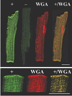
Expression of TASK-1 in rat myocytes.Immunocytochemical staining of rat ventricular myocytes with Anti-KCNK3 (TASK-1) Antibody (#APC-024). A. TASK-1 colocalizes with wheat germ agglutinin (WGA). TASK-1 staining is eradicated when antibody is incubated with the peptide antigen (“-“ panel). B. High resolution of panels in A.Adapted from Jones, S.A. et al. (2002) Am. J. Physiol. 283, 181. with permission of The American Physiological Society.
Applications
Citations
- Human PASMC cell lysate.
Han, L. et al. (2020) Life Sci. 246, 117419. - Rat brain lysate (1:100).
Puissant, M.M. et al. (2017) Front. Cell. Neurosci. 11, 34. - Human atrium lysate (1:200).
Schmidt, C. et al. (2015) Circulation 132, 82.
- Transfected HeLa cells (1:1000).
Renigunta, V. et al. (2014) Mol. Biol. Cell 25, 1877.
- Human bronchial epithelial cells.
Telles, C.J. et al. (2016) Am. J. Physiol. 311, C884. - Rat ventricular myocytes.
Jones, S.A. et al. (2002) Am. J. Physiol. 283, 181.
- Li, M. et al. (2013) J. Biol. Chem. 288, 3668.
- Burdakov, D. et al. (2006) Neuron 50, 711.
- Gonczi, M. et al. (2006) Br. J. Pharmacol. 147, 496.
- Olschewski, A. et al. (2006) Circ. Res. 98, 1072.
- Wareing, M. et al. (2006) Am. J. Physiol. 291, R437.
- Wechselberger, M. et al. (2006) Am. J. Physiol. 291, R518.
- Bai, X. et al. (2005) Reproduction 129, 525.
- Eaton, M.J. et al. (2004) NeuroReport 15, 321.
- Gardener, M.J. et al. (2004) Br. J. Pharmacol. 142, 192.
- Hsu, K. et al. (2004) Molec. Cell 14, 259.
- Lin, W. et al. (2004) J. Neurophysiol. 92, 2909.
- Gurney, A.M. et al. (2003) Circ. Res. 93, 957.
- Cornfield, D.E. et al. (2002) Am. J. Physiol. 283, 1210.
- Hartness, M.E. et al. (2001) J. Biol. Chem. 276, 26499.
- Kindler, C.H. et al. (2000) Brain Res. Mol. Brain. Res. 80, 99.
- Lopes, C.M.B. et al (2000) J. Biol. Chem. 275, 16969.
- Millar, J.A. et al. (2000) Proc. Natl. Acad. Sci. USA 97, 3614.
